. 2018 Feb;22(2):1047-1055.
doi: 10.1111/jcmm.13461.

Study of platelet-rich fibrin combined with rat periodontal ligament stem cells in periodontal tissue regeneration

Affiliations

Study of platelet-rich fibrin combined with rat periodontal ligament stem cells in periodontal tissue regeneration

Xuejing Duan et al. J Cell Mol Med. 2018 Feb.

Abstract

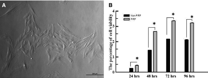
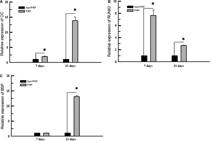
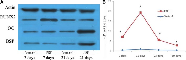
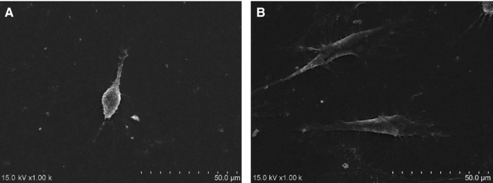
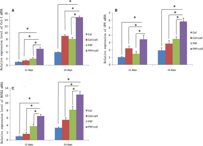
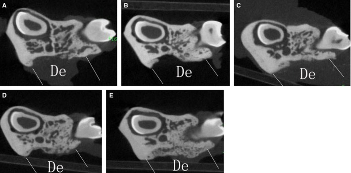
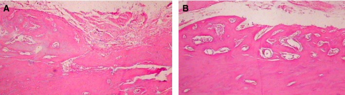
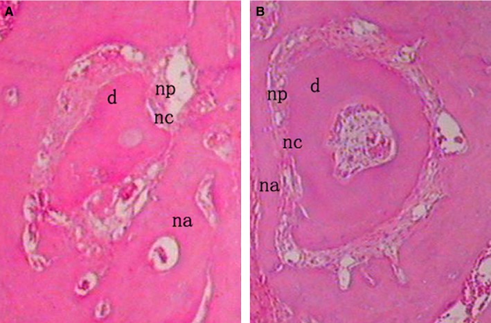
The objective of this study was to investigate the advantages and feasibility of periodontal tissue regeneration using platelet-rich fibrin (PRF) combined with rat periodontal ligament stem cells (PDLSCs) for the first time. We first determined the effect of PRF on rat PDLSCs in vitro. We next conducted an in vivo study, in which a tissue engineering technique was performed to repair periodontal defects in five groups: a blank group, collagen group (implanted collagen membrane), collagen + cells group (implanted collagen membrane and rat PDLSCs), PRF group (implanted PRF membrane) and PRF + cells group (implanted PRF membrane and rat PDLSCs). PRF greatly enhanced cell proliferation, mRNA and protein expression levels of bone sialoprotein (BSP), osteocalcin (OC), and runt-related transcription factor 2 (RUNX2) and activity of alkaline phosphatase (ALP) in vitro. Transplantation of PRF combined with rat PDLSCs resulted in higher expression of osteopontin (Opn), collagen I (COL1A) and RUNX2 at both 12 and 24 days after surgery. Micro-computed tomography and histological analysis showed substantially more new bone formation in the PRF + cells group at 24 days after surgery. Based on these results, we discuss the role of PRF in the proliferation and differentiation of rat PDLSCs and suggest that PRF combined with rat PDLSCs provides a valuable tool for periodontal tissue engineering.

Keywords: periodontal ligament stem cells; periodontal regeneration; platelet-rich fibrin.


Figures

Figure 1
Figure 1
(A) The colonies of rat PDLSCs could be observed microscopically, and cells in good condition exhibited a fusiform shape with an oval nucleus, lying in the middle of the cytoplasm (40×). (B) MTT assay indicated that the proliferation level of rat PDLSCs in PRF group was higher than that in normal group at 24, 48, 72 and 96 hrs in vitro. *Statistically significant difference compared to control and PRF group (P < 0.05).
Figure 2
Figure 2
RT‐PCR was used to measure the mRNA expression levels of BSP, OC and Runx2 of cells cultured in different media after 7 and 14 days. Expressions of OC and Runx2, both significantly increased in PRF group, compared with those exposed to the control group at both 7 and 14 days (A, B). The mRNA expression level of BSP was significantly increased in the cells cultured in the PRF medium at 14 days (C). *Statistically significant difference compared to control and PRF group (P < 0.05).
Figure 3
Figure 3
(A) Western blot analysis was used to measure the protein expression levels of BSP, OC and Runx2 of the cells cultured in different media after 7 and 21 days. The results showed that the protein expression levels of OC and Runx2 were greatly increased in the cells grown in the PRF media at 7 days and those of BSP, OC and Runx2 were all increased at 21 days. (B) ALP activity measurement showed higher ALP activities of rat PDLSCs in the PRF group than those in the control group at 7, 12, 23 and 30 days. *Statistically significant difference compared to control and PRF group (P < 0.05).
Figure 4
Figure 4
(A) Scanning electron microscopy showed that cells cultured in the control medium had a long and thin spindle shape, with tiny, short and less bumps stretching out of the cellular surface. (B) By contrast, cells cultured in the PRF medium were larger and showed a fusiform shape, with radial, longer and many more bumps stretching out of the cellular surface.
Figure 5
Figure 5
RT‐PCR was performed to measure the mRNA expression levels of COL1A, Opn and RUNX2 in different groups. All bone markers showed higher expression levels at 24 days than at 12 days. In addition, the PRF + cells group showed the highest mRNA expression levels of Col1a, Opn and RUNX2 at both 12 and 24 days (A, B, C). *Statistically significant difference (P < 0.05).
Figure 6
Figure 6
Micro‐CT analysis demonstrated that the sample of the PRF + cells group showed much more newly formed hard tissue than those of the other groups at a similar layer and that of the blank group showed the least amount of new bone formation.
Figure 7
Figure 7
(A) At 24 days after surgery, the defect sites in the collagen + cells group were filled with connective tissue and blocked new bone formation at the defect area (100×). (B) In the PRF + cells group, the defect sites exhibited marked bone formation, which almost completely replaced the whole bone defects with new bone spicules (100×).
Figure 8
Figure 8
In the PRF group (A), little new cementum could be observed at the margin of the defect and the PRF + cells group (B) showed a thin layer of new cellular cementum covering the root‐denuded surface at 24 days after surgery. All of the new cementum was cellular cementum, and cracks between the new cementum and the root dentin were observed in some cases. The regenerated PDL fibres separating the new bone from the new cementum was disordered and not perpendicular to the root surface in the PRF + cells group (B) and those in the PRF group (A) were sparse and loose without periodontal fibre bundle formation at 24 days after surgery (×200). d, dentin; np, new periodontal ligament; na, new alveolar bone; nc, new cementum.

References

    1. Ivanovski S. Periodontal regeneration. Aust Dent J. 2009; 54: S118–28. - PubMed
    1. Seo BM, Miura M, Gronthos S, et al Investigation of multipotent postnatal stem cells from human periodontal ligament. Lancet. 2004; 364: 149–55. - PubMed
    1. Liu Y, Zheng Y, Ding G, et al Periodontal ligament stem cell‐mediated treatment for periodontitis in miniature swine. Stem Cells. 2008; 26: 1065–73. - PMC - PubMed
    1. Tassi SA, Sergio NZ, Misawa MYO, et al Efficacy of stem cells on periodontal regeneration: systematic review of pre‐clinical studies. J Periodontal Res. 2017; 52: 793–812. - PubMed
    1. Kim BS, Mooney DJ. Development of biocompatible synthetic extracellular matrices for tissue engineering. Trends Biotechnol. 1998; 16: 224–30. - PubMed

Publication types

MeSH terms