. 2018 Jan;6(2):e13514.
doi: 10.14814/phy2.13514.

Lactobacillus reuteri strains protect epithelial barrier integrity of IPEC-J2 monolayers from the detrimental effect of enterotoxigenic Escherichia coli

Affiliations

Lactobacillus reuteri strains protect epithelial barrier integrity of IPEC-J2 monolayers from the detrimental effect of enterotoxigenic Escherichia coli

Shokoufeh Karimi et al. Physiol Rep. 2018 Jan.

Abstract

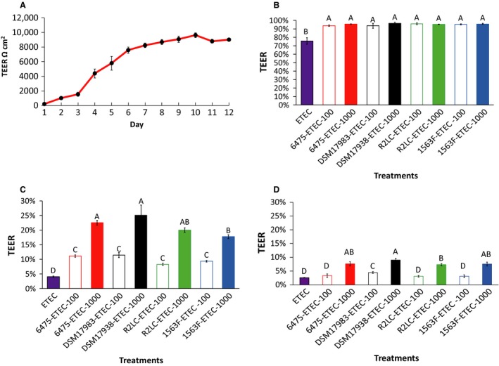
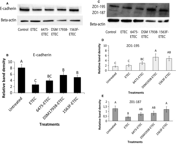
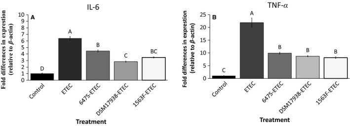
Lactobacillus reuteri is an inhabitant of the gastrointestinal (GI) tract of mammals and birds and several strains of this species are known to be effective probiotics. The mechanisms by which L. reuteri confers its health-promoting effects are far from being fully understood, but protection of the mucosal barrier is thought to be important. Leaky gut is a state of abnormal intestinal permeability with implications for the pathophysiology of various gastrointestinal disorders. Enterotoxigenic Escherichia coli (ETEC) can invade the intestinal mucosa and induce changes in barrier function by producing enterotoxin or by direct invasion of the intestinal epithelium. Our hypothesis was that L. reuteri can protect the mucosal barrier, and the goal of the study was to challenge this hypothesis by monitoring the protective effect of L. reuteri strains on epithelial dysfunction caused by ETEC. Using an infection model based on the porcine intestinal cell line IPEC-J2, it was demonstrated that pretreatment of the cells with human-derived L. reuteri strains (ATCC PTA 6475, DSM 17938 and 1563F) and a rat strain (R2LC) reduced the detrimental effect of ETEC in a dose-dependent manner, as monitored by permeability of FITC-dextran and transepithelial electrical resistance (TEER). Moreover, the results revealed that ETEC upregulated proinflammatory cytokines IL-6 and TNFα and decreased expression of the shorter isoform of ZO-1 (187 kDa) and E-cadherin. In contrast, pretreatment with L. reuteri DSM 17938 and 1563F downregulated expression of IL-6 and TNFα, and led to an increase in production of the longer isoform of ZO-1 (195 kDa) and maintained E-cadherin expression. Interestingly, expression of ZO-1 (187 kDa) was preserved only when the infected cells were pretreated with strain 1563F. These findings demonstrate that L. reuteri strains exert a protective effect against ETEC-induced mucosal integrity disruption.

Keywords: Lactobacillus reuteri; Enterotoxigenic Escherichia coli (ETEC); IPEC-J2; mucosal integrity.


Figures

Figure 1
Figure 1
Effect of Lactobacillus reuteri strains on the deleterious effect of ETEC on transepithelial electrical resistance (TEER). The IPEC‐J2 cells were grown on transwell filters for 12 days and TEER was measured every day. A representative experiment of progression of TEER values (A). Polarized monolayers were pretreated or not pretreated with L. reuteri strains ATCC PTA 6475, DSM 17938, R2LC and 1563F at multiplicity of bacteria (MOB) of 100:1 and 1000:1 for 6 h. After 6 h exposure, L. reuteri was washed off and the monolayer was infected with ETEC at multiplicity of infection (MOI) 10 for 6 h. TEER measurement, 2 h postinfection (B). TEER measurement, 4 h postinfection (C). TEER measurement, 6 h postinfection (D). TEER was measured in ohms and corrected for the resistance of blank filters and for the membrane area and expressed as percentage of the starting value. Data are given as means (±SEM) three‐four independent seedings. Columns with different letters are significantly different (P ≤ 0.05).
Figure 2
Figure 2
Effect of Lactobacillus reuteri strains on FITC‐dextran permeability enhanced by ETEC. Monolayers were pretreated with four strains of L. reuteri at MOB 100 and 1000 for 6 h, half of them were challenged with ETEC for 6 h, and finally 1 mg/mL of FITC‐dextran (4 kDa) was added to the apical side of the inserts. The samples were taken from the basolateral pole 6 h post‐treatment and analysed for fluorescence intensity (excitation, 492 nm; emission, 520 nm). Data given are means (±SEM) of three independent seedings. Values with different letters are significantly different at  0.05.
Figure 3
Figure 3
Effect of Lactobacillus reuteri strains and ETEC on tight junction protein expression in IPEC‐J2 cells. Cells were grown in six‐well culture plates to a density of 1.5 × 106 cells/well. Polarised monolayers were pretreated with L. reuteri strains ATCC PTA 6475, DSM 17938 and 1563F (MOB 100:1) for 6 h and then the monolayers were infected with ETEC (MOI 10:1) for 4 h. The immunoblotting experiments were performed using 40 μg of IPEC‐J2 protein per well. A band of 135 kDa corresponding to E‐cadherin (A). Two bands of 195 kDa and 187 kDa, respectively, corresponding to two different isoforms of ZO‐1, appeared (C). Relative protein levels were quantified using densitometry and expressed as optical density ratio: Relative band densities are shown for E‐cadherin (B), ZO‐1 (195 kDa) (D) and ZO‐1 (187 kDa) (E). The immunoblotting experiments were performed on four independent seedings and the densitometric values of ZO‐1 and E‐cadherin normalized to reference protein, with β‐actin as internal standard. Values are mean ± SEM. Columns with different letters are significantly different (P ≤ 0.05) as determined by ANOVA.
Figure 4
Figure 4
Effect of Lactobacillus reuteri strains on ETEC‐induced IL‐6 and TNF α expression in IPEC‐J2 cells. IL‐6 expression (A) and TNF α expression (B). Cells were grown in six‐well tissue culture plates to a density of 1.5 × 106. Polarized monolayers were pretreated with L. reuteri strains ATCC PTA 6475, DSM 17938 and 1563F (MOB 100:1) for 6 h. Thereafter, monolayers were challenged with ETEC (MOI 10:1) for 4 h. The results were considered significant at P < 0.05, as determined by ANOVA. The data represent results from 3 to 4 independent seedings.

Similar articles

Cited by

References

    1. Ahrné, S. , Molin G., and Axelsson L.. 1992. Transformation of Lactobacillus reuteri with electroporation: studies on the erythromycin resistance plasmid pLUL631. Curr. Microbiol. 24:199–205.
    1. Al‐Sadi, R. , Boivin M., and Ma T.. 2009. Mechanism of cytokine modulation of epithelial tight junction barrier. Front. Biosci. 14:2765–2778. - PMC - PubMed
    1. Al‐Sadi, R. , Ye D., Boivin M., Guo S., Hashimi M., Ereifej L., et al. 2014. Interleukin‐6 modulation of intestinal epithelial tight junction permeability is mediated by JNK pathway activation of Claudin‐2 Gene. PLoS ONE 9:e85345. - PMC - PubMed
    1. Anderson, R. C. , Cookson A. L., McNabb W. C., Park Z., McCann M. J., Kelly W. J., et al. 2010. Lactobacillus plantarum MB452 enhances the function of the intestinal barrier by increasing the expression levels of genes involved in tight junction formation. BMC Microbiol. 10:316. - PMC - PubMed
    1. Assimakopoulos, S. F. , Papageorgiou I., and Charonis A.. 2011. Enterocytes’ tight junctions: from molecules to diseases. World J. Gastrointest. Pathophysiol. 2:123–137. - PMC - PubMed

Publication types