. 2018 Jan 24;15(1):22.
doi: 10.1186/s12985-018-0930-x.

Ubiquitous giants: a plethora of giant viruses found in Brazil and Antarctica

Affiliations

Ubiquitous giants: a plethora of giant viruses found in Brazil and Antarctica

Ana Cláudia Dos S P Andrade et al. Virol J. .

Abstract

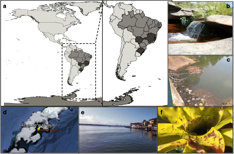
Background: Since the discovery of giant viruses infecting amoebae in 2003, many dogmas of virology have been revised and the search for these viruses has been intensified. Over the last few years, several new groups of these viruses have been discovered in various types of samples and environments.In this work, we describe the isolation of 68 giant viruses of amoeba obtained from environmental samples from Brazil and Antarctica.

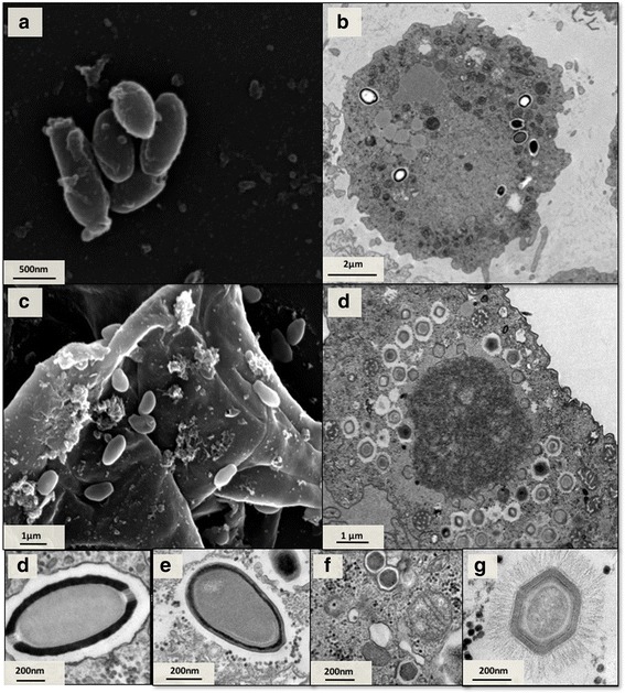
Methods: Isolated viruses were identified by hemacolor staining, PCR assays and electron microscopy (scanning and/or transmission).

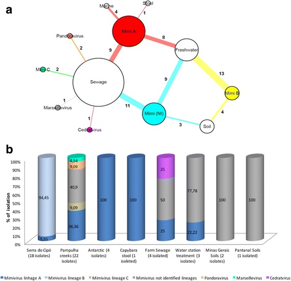
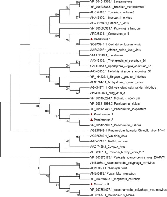
Results: A total of 64 viruses belonging to the Mimiviridae family were isolated (26 from lineage A, 13 from lineage B, 2 from lineage C and 23 from unidentified lineages) from different types of samples, including marine water from Antarctica, thus being the first mimiviruses isolated in this extreme environment to date. Furthermore, a marseillevirus was isolated from sewage samples along with two pandoraviruses and a cedratvirus (the third to be isolated in the world so far).

Conclusions: Considering the different type of samples, we found a higher number of viral groups in sewage samples. Our results reinforce the importance of prospective studies in different environmental samples, therefore improving our comprehension about the circulation anddiversity of these viruses in nature.

Keywords: Antarctica; Brazil; Cedratvirus; Giant viruses; Marseillevirus; Mimivirus; Pandoravirus; Prospection.


Conflict of interest statement

Ethics approval and consent to participate

Two hundred samples of human nasopharyngeal aspiratewere also kindly provided by Laboratório Central do Estado do Rio Grande do Sul (LACEN/RS) (Table 1). These samples were used underapproval of the ethics committee of Universidade Federal de Ciências da Saúde de Porto Alegre (protocol number 1774/12, register 928/12).

Consent for publication

Not applicable

Competing interests

The authors declare that they have no competing interests.

Publisher’s Note

Springer Nature remains neutral with regard to jurisdictional claims in published maps and institutional affiliations.

Figures

Fig. 1
Fig. 1
Locations where the environmental samples were collected. Schematic map (a) indicating in dark gray the location of collections tested and pictures from representative areas represented by letters (b-f). River at Serra do Cipó, MG, Brazil (b); Sewage creeks, Belo Horizonte, MG, Brazil (c); Location of collection of one of marine water samples in Antarctica (d); Location of collection of mangrove water, ES, Brazil (e). Bromeliads at Serra da Saudinha, AL, Brazil (f)
Fig. 2
Fig. 2
Diversity of isolated giant virus by type of sample and collections. Network graph showing the viral groups isolated and identified by PCR and electron microscopy assays in different samples. Each node represents a type of sample (white nodes) or viral group (colored nodes). The node diameter is proportional to the edge degree. The numbers of isolated viruses in each sample are shown on the respective edge. The layout was generated using a force based algorithm followed by manual rearrangement for a better visualization of the connections (a). A total of 7 viral groups are represented. Isolation rate of each virus groups by collections (b)
Fig. 3
Fig. 3
Phylogenetic tree of isolates. A neighbor-joining phylogenetic tree constructed using a 327 amino acid fragment of the DNA polymerase B gene. Tree was constructed by using MEGA version 7.0 (www.megasoftware.net) on the basis of the amino acids sequences with 1000 bootstrap replicates. Bootstrap values > 40% are shown. Nucleotide sequences were obtained from GenBank. The isolates are highlighted with red triangle. Scale bar indicates rate of evolution
Fig. 4
Fig. 4
Electron microscopy images of viruses isolated. SEM of Cedratvirus isolated from sewage farm of MG (a) TEM (b) and SEM (c) of Pandoravirus isolate from Mergulhão sewage creek. TEM of Mimivirus isolated from Antarctica (c) TEM of Cedratvirus isolated from sewage farm of MG (d) TEM of Pandoravirus isolate from Bom Jesus sewage creek (e) marseillevirus isolated from Bom Jesus sewage creek (f). TEM mimivirus particle detail that was isolated from Antarctica (g). Scale Bars: (a-d) 500 nm; (e) 50 nm

References

    1. La Scola B, Audic S, Robert C, Jungang L, de Lamballerie X, Drancourt M, et al. A giant virus in amoebae. Science. 2003;299:2033. doi: 10.1126/science.1081867. - DOI - PubMed
    1. Aherfi S, Colson P, La Scola B, Raoult D. Giant viruses of amoebas: an update. Front Microbiol. 2016;7:1–14. doi: 10.3389/fmicb.2016.00349. - DOI - PMC - PubMed
    1. Boyer M, Yutin N, Pagnier I, Barrassi L, Fournous G, Espinosa L, et al. Giant Marseillevirus highlights the role of amoebae as a melting pot in emergence of chimeric microorganisms. Proc Natl Acad Sci. 2009;106:21848–21853. doi: 10.1073/pnas.0911354106. - DOI - PMC - PubMed
    1. Legendre M, Bartoli J, Shmakova L, Jeudy S, Labadie K, Adrait A, et al. Thirty-thousand-year-old distant relative of giant icosahedral DNA viruses with a pandoravirus morphology. Proc Natl Acad Sci. 2014;111:4274–4279. doi: 10.1073/pnas.1320670111. - DOI - PMC - PubMed
    1. Abrahão JS, Araújo R, Colson P, La Scola B. The analysis of translation-related gene set boosts debates around origin and evolution of mimiviruses. PLoS Genet. 2017;16:1–12. - PMC - PubMed

Publication types

LinkOut - more resources