. 2018 Jan 25;13(1):e0191617.
doi: 10.1371/journal.pone.0191617. eCollection 2018.

Inhibition of EV71 by curcumin in intestinal epithelial cells

Affiliations

Inhibition of EV71 by curcumin in intestinal epithelial cells

Hsing-I Huang et al. PLoS One. .

Abstract

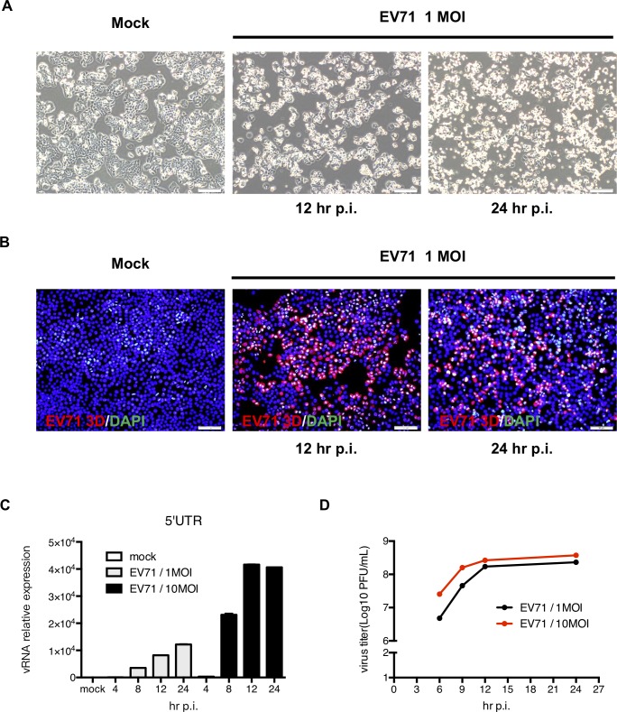
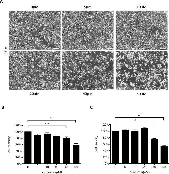
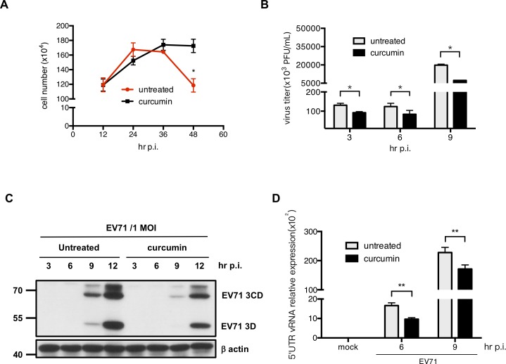
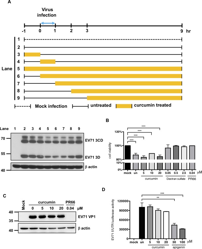
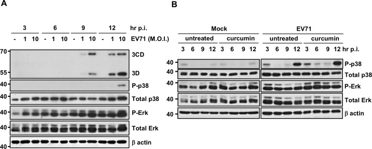
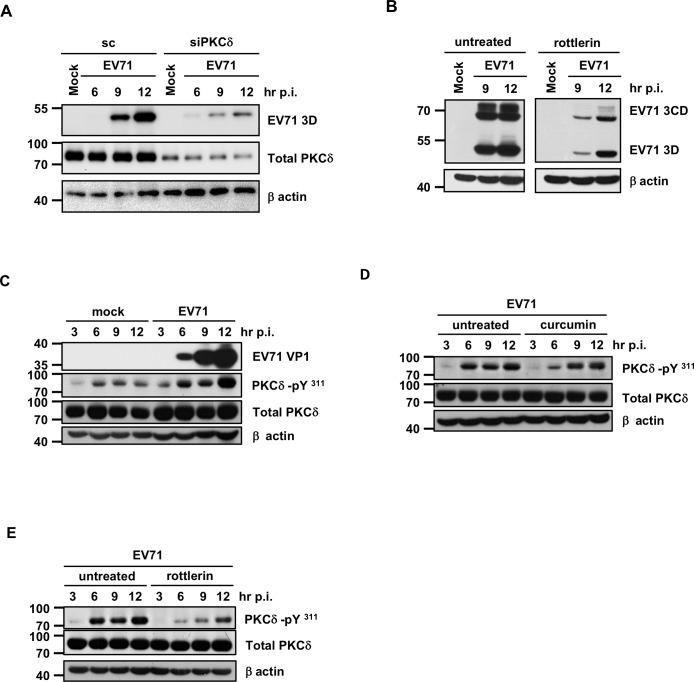
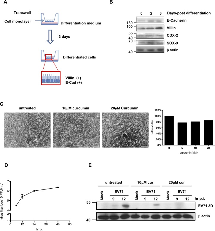
EV71 is a positive-sense single-stranded RNA virus that belongs to the Picornaviridae family. EV71 infection may cause various symptoms ranging from hand-foot-and-mouth disease to neurological pathological conditions such as aseptic meningitis, ataxia, and acute transverse myelitis. There is currently no effective treatment or vaccine available. Various compounds have been examined for their ability to restrict EV71 replication. However, most experiments have been performed in rhabdomyosarcoma or Vero cells. Since the gastrointestinal tract is the entry site for this pathogen, we anticipated that orally ingested agents may exert beneficial effects by decreasing virus replication in intestinal epithelial cells. In this study, curcumin (diferuloylmethane, C21H20O6), an active ingredient of turmeric (Curcuma longa Linn) with anti-cancer properties, was investigated for its anti-enterovirus activity. We demonstrate that curcumin treatment inhibits viral translation and increases host cell viability. Curcumin does not exert its anti-EV71 effects by modulating virus attachment or virus internal ribosome entry site (IRES) activity. Furthermore, curcumin-mediated regulation of mitogen-activated protein kinase (MAPK) signaling pathways is not involved. We found that protein kinase C delta (PKCδ) plays a role in virus translation in EV71-infected intestinal epithelial cells and that curcumin treatment decreases the phosphorylation of this enzyme. In addition, we show evidence that curcumin also limits viral translation in differentiated human intestinal epithelial cells. In summary, our data demonstrate the anti-EV71 properties of curcumin, suggesting that ingestion of this phytochemical may protect against enteroviral infections.


Conflict of interest statement

Competing Interests: The authors have declared that no competing interests exist.

Figures

Fig 1
Fig 1. HT-29 cells are permissive to EV71 infection.
(A) HT29 cells were seeded on culture plates and infected with EV71 at the MOI of 1. Cell morphology was observed using an inverted microscope (magnification = 200x). (B) To confirm infection, the cells were fixed and reacted with a primary anti-EV71 3D antibody. A PE-conjugated anti-mouse IgG antibody was then applied. DAPI was used to stain the cell nuclei (magnification = 200x). (C) Total RNA was extracted from mock-infected and EV71-infected cells, and RT-qPCR was performed to detect the quantity of viral RNA. (D) Total cell lysates were harvested to detect the viral titers using a plaque assay.
Fig 2
Fig 2. Curcumin cytotoxicity assay.
(A) To detect the cytotoxicity of curcumin in host cells, HT29 cells were seeded and treated with various concentrations of curcumin for 2 days. Cell morphology was observed and recorded (magnification = 200x). (B) Trypan blue exclusion and (C) MTT assays were performed to determine the viability of treated cells.
Fig 3
Fig 3. Curcumin treatment increases the survival of host cells and suppresses EV71 replication.
(A) Cells were seeded in culture plates and treated with 10 μM curcumin. After one hour, the medium was removed, and the cells were infected with EV71 at the MOI of 1 in curcumin-containing serum-free medium. After adsorption, the medium was removed, and medium containing 2% serum and 10 μM curcumin was added. The growth curves of infected cells were counted using a trypan blue exclusion assay. (B) Cell lysates were collected from cell samples, and a plaque assay was performed to determine the virus titers. (C) Cells were harvested at different time points, and total protein was extracted to determine the expression of EV71 3D protein by Western blot. The expression of β-actin was used as an internal control. (D) RT-qPCR analysis was performed to detect the amounts of viral RNA.
Fig 4
Fig 4. Curcumin inhibits EV71 protein expression when added at the early stages of infection.
(A) A time-of-addition assay was performed to assess the anti-EV71 mechanism of curcumin. HT29 cells were seeded on culture plates and infected with EV71 at the MOI of 1. Viral adsorption was allowed to occur from 0 to 1 hour p.i. Curcumin (10 μM) was added to the cells at the indicated times. After treatment, the cells were washed three times, and the medium was replaced. After 9 hours of infection, the cells were harvested, and total protein was isolated for Western blot analysis. An anti-EV71 3D antibody was used to detect the viral protein expression level. Expression of β-actin was used as an internal control. (B) To test whether curcumin can modulate the virus adsorption to the cells, EV71 virus infected cells in the absence or presence of curcumin, dextran sulfate or PR66 for one hour on ice. And cell survival rates were determined at 48 hours p.i. by MTT assay. (C) To test whether curcumin can modulate the EV71 viral particles binding to cells, EV71 were incubated with different concentrations of curcumin, or PR66 at 0.04 μM for 1 hour on ice and then used to infect the HT29 cells. Total protein was extracted at 12 hours p.i. and subsequently subjected for Western blot to examine the expression levels of viral protein VP1 and host β-actin. (D) To test whether curcumin could modulate the activity of EV71 5’UTR IRES, in vitro translation assay was used to detect the EV71 5’UTR IRES luciferase activity in the absence or presence of curcumin or apigenin. Un: untreated.
Fig 5
Fig 5. Curcumin does not suppress EV71 by modifying MAPK pathways.
(A) Cells were infected with EV71 at different MOIs. Total cell lysates were harvested at different time points, and Erk and p38 phosphorylation was detected by immunoblot analysis. (B) To detect the effect of curcumin on MAPK pathways, HT29 cells were seeded in plates and infected with EV71 at the MOI of 1 with or without 10 μM curcumin in the medium. The cells were harvested at different time points, and total protein was isolated for Western blot analysis. Anti-P-Erk, anti-P-p38, anti-Total Erk, anti-Total p38, and anti-EV71 3D antibodies were applied to detect the phosphorylation of kinases.
Fig 6
Fig 6. Curcumin inhibits EV71 infection-induced PKCδ phosphorylation.
(A) PKCδ siRNA and scramble siRNA were transfected into HT29 cells for 24 hours, and the cells were then infected with EV71 at the MOI of 1. Total protein samples were isolated at different time points and subjected to Western blot analysis to examine the expression levels of PKCδ and EV71 viral protein 3D. (B) HT29 cells were seeded and infected with EV71 in the presence of rottlerin (5 μM), a known PKCδ inhibitor. The expression levels of viral protein 3D and β-actin were then analyzed by Western blot. (C) Protein samples were harvested from mock-infected and EV71-infected HT29 cells at various time points. Immunoblotting was performed to examine the expression levels of viral protein VP1, total PKCδ, P-PKCδ (Tyr311) and β-actin. (D) Total protein was extracted from EV71-infected HT29 cells in the presence or absence of 10 μM curcumin. The expression of PKCδ, P-PKCδ (Tyr311), EV71 3D, and β-actin was analyzed by Western blot. (E) Cells were treated with rottlerin and infected with EV71. Western blot analysis was performed to detect the expression levels of P-PKCδ (Tyr311), Total PKCδ and β-actin.
Fig 7
Fig 7. Curcumin treatment suppresses EV71 translation in differentiated C2BBe1 cells.
(A) C2BBel cells were seeded on transwell plates and cultivated in enterocyte differentiation medium for 3 days. Total protein was isolated from cells at different differentiation time points. (B) Western blot analysis was performed to measure the expression levels of E-cadherin, Villin, CDX-2, and SOX-9. The expression of β-actin was used as an internal control. (C) Differentiated C2BBe1 cells were treated with 10 and 20 μM curcumin for 48 hours and the morphologies were observed using inverted microscope (Magnification = 200x). Trypan blue was added and the cell viabilities were counted. (D) Differentiated C2BBe1 cells were seeded and infected with EV71 at the MOI of 2. Total cell lysates were collected at different time points and subjected to plaque assays to determine the viral titers. (E) Differentiated C2BBe1 cells were infected with EV71 at the MOI of 10 in the absence or presence of curcumin (10 and 20 μM). The cells were harvested at different time points for protein isolation. The expression levels of viral protein 3D were measured by Western blot analysis.

References

    1. Chan KP, Goh KT, Chong CY, Teo ES, Lau G, Ling AE. Epidemic hand, foot and mouth disease caused by human enterovirus 71, Singapore. Emerg Infect Dis. 2003; 9: 78–85. doi: 10.3201/eid1301.020112 - DOI - PMC - PubMed
    1. Shimizu H, Utama A, Onnimala N, Li C, Li-Bi Z, Yu-Jie M et al. Molecular epidemiology of enterovirus 71 infection in the Western Pacific Region. Pediatr Int. 2004; 46: 231–235. doi: 10.1046/j.1442-200x.2004.01868.x - DOI - PubMed
    1. Chan LG, Parashar UD, Lye MS, Ong FG, Zaki SR, Alexander JP et al. Deaths of children during an outbreak of hand, foot, and mouth disease in sarawak, malaysia: clinical and pathological characteristics of the disease. For the Outbreak Study Group. Clin Infect Dis. 2000; 31: 678–683. doi: 10.1086/314032 - DOI - PubMed
    1. Galasiti Kankanamalage AC, Kim Y, Weerawarna PM, Uy RA, Damalanka VC, Mandadapu SR et al. Structure-guided design and optimization of dipeptidyl inhibitors of norovirus 3CL protease. Structure-activity relationships and biochemical, X-ray crystallographic, cell-based, and in vivo studies. J Med Chem. 2015; 58: 3144–3155. doi: 10.1021/jm5019934 - DOI - PMC - PubMed
    1. Yang Y, Xiu J, Liu J, Zhang L, Li X, Xu Y et al. Chebulagic acid, a hydrolyzable tannin, exhibited antiviral activity in vitro and in vivo against human enterovirus 71. Int J Mol Sci. 2013; 14:9618–9627. doi: 10.3390/ijms14059618 - DOI - PMC - PubMed

Publication types

MeSH terms

LinkOut - more resources