. 2018 Jan 25;14(1):e1006838.
doi: 10.1371/journal.ppat.1006838. eCollection 2018 Jan.

MERS-CoV 4b protein interferes with the NF-κB-dependent innate immune response during infection

Affiliations

MERS-CoV 4b protein interferes with the NF-κB-dependent innate immune response during infection

Javier Canton et al. PLoS Pathog. .

Abstract

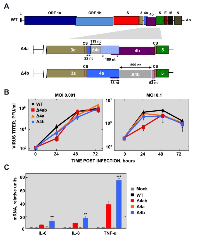
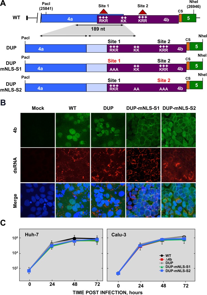
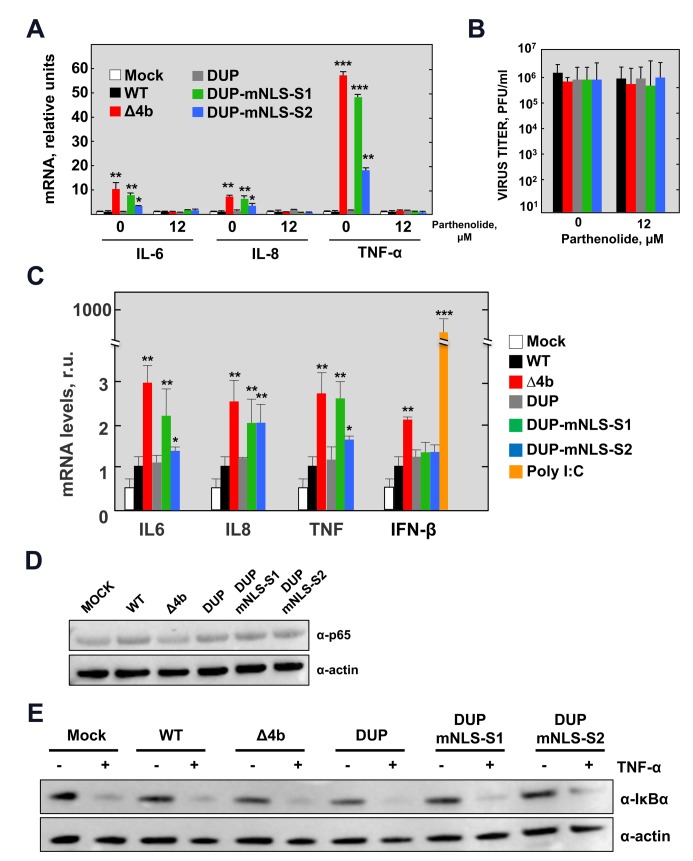
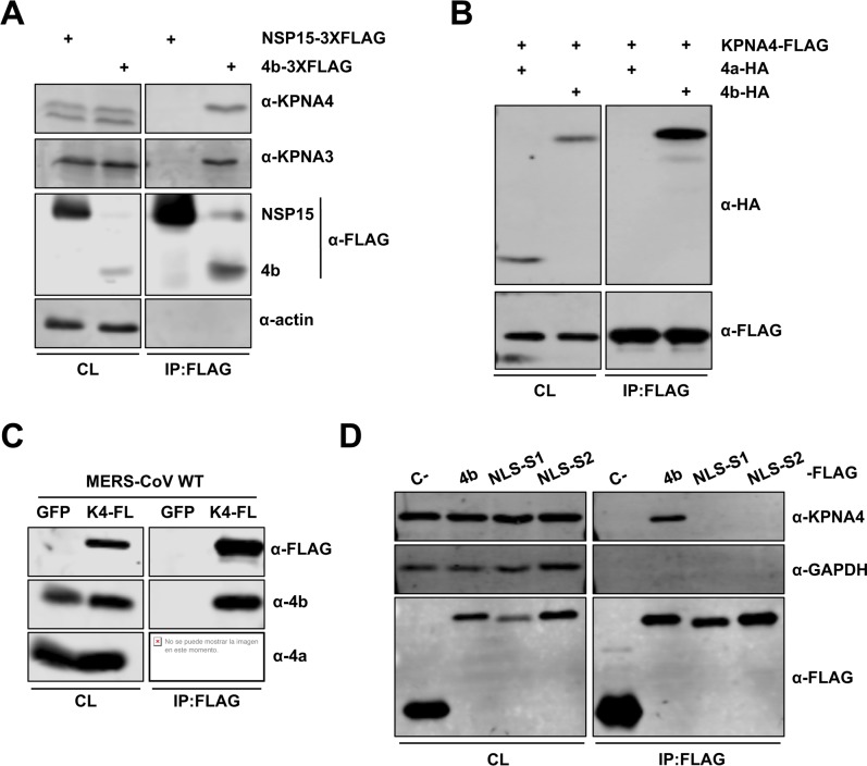
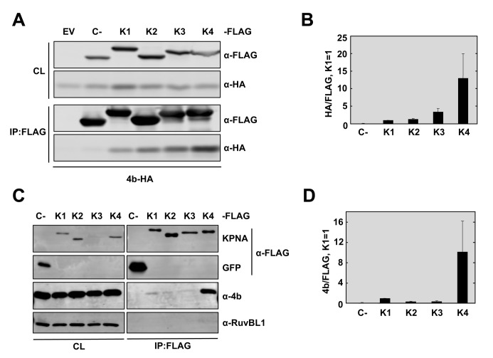
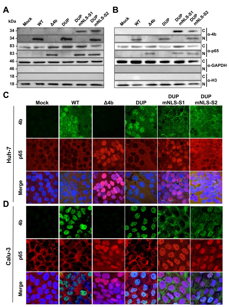
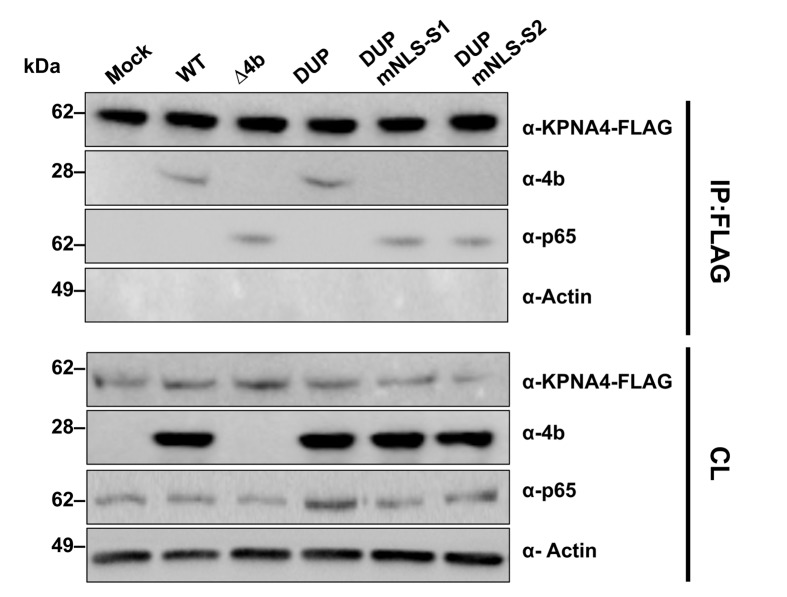
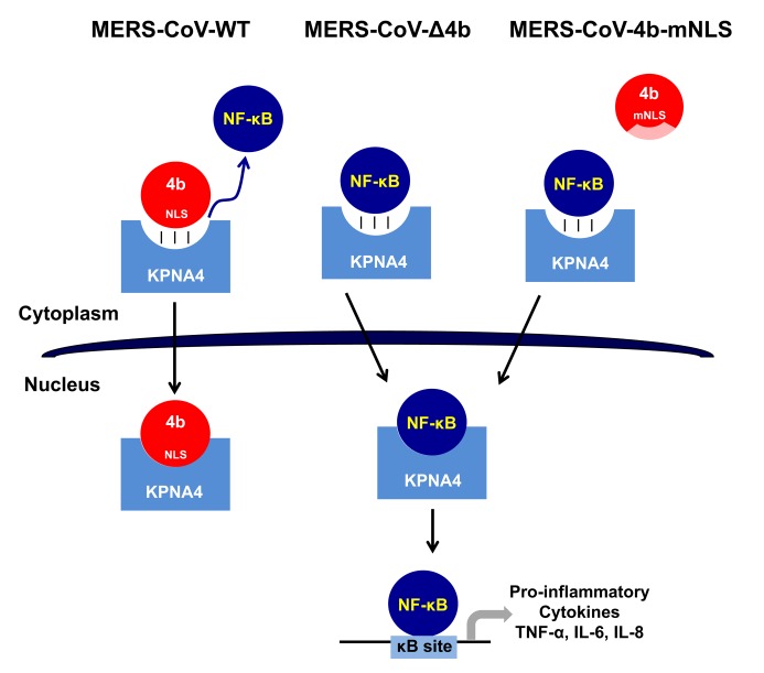
Middle East respiratory syndrome coronavirus (MERS-CoV) is a novel human coronavirus that emerged in 2012, causing severe pneumonia and acute respiratory distress syndrome (ARDS), with a case fatality rate of ~36%. When expressed in isolation, CoV accessory proteins have been shown to interfere with innate antiviral signaling pathways. However, there is limited information on the specific contribution of MERS-CoV accessory protein 4b to the repression of the innate antiviral response in the context of infection. We found that MERS-CoV 4b was required to prevent a robust NF-κB dependent response during infection. In wild-type virus infected cells, 4b localized to the nucleus, while NF-κB was retained in the cytoplasm. In contrast, in the absence of 4b or in the presence of cytoplasmic 4b mutants lacking a nuclear localization signal (NLS), NF-κB was translocated to the nucleus leading to the expression of pro-inflammatory cytokines. This indicates that NF-κB repression required the nuclear import of 4b mediated by a specific NLS. Interestingly, we also found that both in isolation and during infection, 4b interacted with α-karyopherin proteins in an NLS-dependent manner. In particular, 4b had a strong preference for binding karyopherin-α4 (KPNA4), which is known to translocate the NF-κB protein complex into the nucleus. Binding of 4b to KPNA4 during infection inhibited its interaction with NF-κB-p65 subunit. Thereby we propose a model where 4b outcompetes NF-κB for KPNA4 binding and translocation into the nucleus as a mechanism of interference with the NF-κB-mediated innate immune response.


Conflict of interest statement

The authors have declared that no competing interests exist.

Figures

Fig 1
Fig 1. Generation of MERS-CoV-Δ4a and Δ4b deletion mutants.
(A) Schematic representation of MERS-CoV WT genome. Essential viral genes (ORF1a, ORF1b, S, E, M, N) and accessory genes (3, 4a, 4b and 5) are indicated. L, leader sequence; An, poly(A) tail; CS, conserved sequence included in transcription-regulating sequences. Genes 4a and 4b were deleted from the MERS-CoV infectious cDNA clone by PCR-directed mutagenesis as described in Materials and Methods. Deleted regions within 4a and 4b genes are illustrated as light grey boxes. The size of both deleted and remaining regions in 4a and 4b genes is indicated by arrows. The patterned boxes in Δ4a and Δ4b mutants indicate frameshift mutations in 4a and 4b sequence caused by the deletions. (B) Growth kinetics of Δ4a and Δ4b deletion mutants in Huh-7 cells at the indicated MOIs. Supernatants were collected at 24, 48 and 72 hpi and titrated by plaque assay. (C) NF-κB-dependent cytokine response of Huh-7 cells either mock-infected or infected with WT, Δ4ab, Δ4a or Δ4b mutants (MOI = 1 PFU/cell). At 24 hpi, mRNA levels of IL-6, IL-8 and TNF-α were quantified by RT-qPCR and compared to those in WT-infected cells, using the ΔΔCt method for calculation and HMBS as a reference endogenous gene. Shown are means with standard deviations, which were analyzed using an unpaired t-test against the wild-type (**, p<0.01; ***, p<0.001).
Fig 2
Fig 2. Generation of MERS-CoV-4b-NLS mutants.
(A) Schematic representation of 4a and 4b overlapping sequences in MERS-CoV WT (top) and 4b-NLS mutants (bottom). Nuclear localization signals in 4b protein (NLS-S1 and NLS-S2), consisting of positively charged amino acids lysine (K) and arginine (R) are indicated. The 189 nt-sequence duplicated in 4b-NLS mutants is indicated by the shadowed area and the arrows. DUP, mutant including WT duplicated 4a-4b sequences, used as a control; DUP-mNLS-S1, mutant with 4b NLS Site 1 mutated to alanine; DUP-mNLS-S2, mutant with Site 2 and two close Lys residues mutated to alanine, as described in Materials and Methods. The light blue boxes in DUP, DUP-mNLS-S1 and DUP-mNLS-S2 mutants indicate duplicated 4a sequences. PacI and NheI restriction sites used for the assembly of infectious cDNA clones and their genomic positions (first nucleotide of the recognition sequence) are indicated. (B) Analysis by confocal microscopy of 4b subcellular localization in Huh-7 cells either mock-infected or infected with MERS-CoV-4b-NLS mutants (MOI = 0.1 PFU/cell, 24 hpi). 4b protein (green) and dsRNA (red) were detected with specific antibodies, while nuclei (blue) were stained with DAPI. (C) Growth kinetics of MERS-CoV-4b-NLS mutants in Huh-7 and Calu-3 cells at an MOI of 0.1. Supernatants were collected at 24, 48 and 72 hpi and titrated by plaque assay. Error bars represent SD.
Fig 3
Fig 3. NF-κB-dependent cytokine response during infection with MERS-CoV-4b-NLS mutants (MOI = 1 PFU/cell, 24 hpi).
(A) The mRNA expression levels of IL-6, IL-8 and TNF-α were quantified by RT-qPCR in Huh-7 cells either mock-treated or treated with the NF-κB inhibitor parthenolide (12 μM), as described in Fig 1. Error bars represent SD. (B) Viral titers in the supernatant of Huh-7 cells infected with MERS-CoV-4b-NLS mutants either mock-treated or treated with parthenolide. (C) The mRNA expression levels of IL6, IL8, TNF-α and IFNB1 were quantified by RT-qPCR in Calu-3 cells infected with MERS-CoV-4b-NLS mutants as described in S1 Fig. (D) Analysis by Western-blot of NF-κB p65 levels at 24 hpi in Huh-7 cells infected with 4b-NLS mutants (MOI = 1 PFU/cell). (E) Analysis by Western-blot of TNF-α induced IκBα degradation in Huh-7 cells either mock-infected or infected with MERS.CoV-4b-NLS mutants (MOI = 1 PFU/cell). At 14 hpi, cell supernatant was replaced by fresh medium containing TNF-α (50 ng/ml). After 30 min treatment, cell lysates were prepared for immunoblotting with anti IκBα antibody. Actin was used as a loading control. Shown are means with standard deviations, which were analyzed using an unpaired t-test against the wild-type (*, p<0.1**; p<0.01; ***, p<0.001).
Fig 4
Fig 4. MERS-CoV 4b protein interacts with importin-α2 family karyopherins.
(A-B). 4b interacts with KPNA3 and KPNA4 when expressed in isolation. Huh-7 cells were transfected with plasmids expressing NSP15-3XFLAG or 4b-3XFLAG (A) or KPNA4-FLAG and 4a-HA or 4b-HA (B). Cells were collected 48 hours later and cell lysates were immunoprecipitated with anti-FLAG monoclonal antibody. Cell lysates (CL) and eluted proteins were analyzed by immunoblotting with indicated antibodies. (C) 4b interacts with KPNA4 during infection. Huh-7 cells were transfected with plasmids expressing GFP or KPNA4-FLAG (K4-FL). 48 hours later cells were infected with MERS-CoV at an MOI of 0.1 PFU/cell. Cells were collected at 20 hpi and cell lysates were immunoprecipitated and analyzed as described above. (D) 4b requires both NLS Site 1 and Site 2 for efficient interaction with KPNA4. Huh-7 cells were transfected with plasmids expressing sMacro-3XFLAG (C-), 4b-3XFLAG (4b), or the 4b-NLS mutants 4b-mNLS-S1–3XFLAG (NLS-S1), or 4b-mNLS-S2–3XFLAG (NLS-S2). Cells were collected 48 hours later and cell lysates were immunoprecipitated and analyzed as described above. Viral protein 4a and cell proteins actin and GAPDH have been used as controls for non-specific binding to KPNA4-FLAG or to FLAG-antibody coated beads. GFP has been used as a control for 4b non-nonspecific binding to the FLAG-antibody coated beads in the absence of karyopherin protein.
Fig 5
Fig 5. MERS-CoV 4b has higher affinity for interaction with karyopherin-α4 than other karyopherins.
(A) Huh-7 cells were co-transfected with plasmids expressing 4b-HA and empty vector (EV) or Control-3XFLAG (C-) or KPNA-FLAG proteins KPNA1-FLAG (K1), KPNA2-FLAG (K2), KPNA3-FLAG (K3), or KPNA4-FLAG (K4). Cells were collected 48 hours later and cell lysates (CL) were immunoprecipitated with anti-FLAG monoclonal antibody. Cell lysates and eluted proteins were analyzed by immunoblotting with indicated antibodies. (B) Quantification of 4b-karyopherin interactions. The relative binding of 4b to each karyopherin was determined by measuring the HA signal and dividing by the FLAG signal, derived using Image Studio Software. The ratio of 4b-HA to KPNA1-FLAG was set to 1. Data represent the combined results of 2 independent experiments. Error bars represent SD. (C) Huh-7 cells were transfected with plasmids expressing GFP-3XFLAG (C-) or KPNA-FLAG proteins KPNA1-FL (K1), KPNA2-FL (K2), KPNA3-FL (K3), or KPNA4-FL (K4). 48 hours later, cells were infected with WT MERS-CoV at a MOI of 0.1 PFU/cell. Cells were collected at 20 hpi and cell lysates were immunoprecipitated with anti-FLAG monoclonal antibody and analyzed as described in (A). Cell protein RuvBL1 has been used as a control for non-specific binding to KPNA4-FLAG or to FLAG-antibody coated beads. GFP and C- have been used as controls for 4b non-nonspecific binding to the FLAG-antibody coated beads in the absence of karyopherin protein. (D) The relative binding of 4b to each karyopherin during infection was determined as described in (B). These data represent the combined results of 2 independent experiments. Error bars represent SD.
Fig 6
Fig 6. Subcellular localization of NF-κB during infection with MERS-CoV 4b-NLS mutants.
Huh-7 (A) or Calu-3 (B) cells were mock-infected or infected (MOI = 0.1 PFU/cell) with WT, Δ4b or 4b-NLS mutants. At 18 hpi, cells were fractionated into cytoplasmic (C) and nuclear (N) fractions and analyzed by Western-blot for 4b and p65 detection. GAPDH and histone H3 were used as cytoplasmic and nuclear markers, respectively. Huh-7 (C) or Calu-3 (D) cells were infected with WT, Δ4b or 4b-NLS mutants (MOI 0.1 PFU/cell). At 24 hpi, cells were fixed and stained with antibodies against 4b (green) and p65 (red). Cell nuclei were stained with DAPI (blue).
Fig 7
Fig 7. Interference of MERS-CoV 4b protein in NF-κB-karyopherin 4 binding during infection.
Huh-7 cells were transfected with a plasmid encoding karyopherin 4 (KPNA4)-FLAG. At 24 hpt, cells were infected (MOI 0.1 PFU/cell) with WT, Δ4b or 4b-NLS mutants. At 20 hpi, cells lysates were immunoprecipitated with anti-FLAG antibodies. Cell lysates (CL) and eluted proteins were analyzed by immunoblotting with indicated antibodies.
Fig 8
Fig 8. Proposed mechanism of 4b action in MERS-CoV infection.
In WT MERS-CoV infection, 4b protein with an intact NLS binds KPNA4 for nuclear translocation thereby outcompeting NF-κB binding to KPNA4, which retains NF-κB in the cytoplasm and inhibits the expression of NF-κB-dependent pro-inflammatory cytokines. In contrast, in the absence of 4b (MERS-CoV-Δ4b) or in the presence of cytoplasmic 4b NLS-mutants (MERS-CoV-4b-mNLS), which do not bind KPNA4, NF-κB is imported into the nucleus by KPNA4, where it binds to DNA κB sites to promote the expression of pro-inflammatory cytokines (TNF-α, IL-6 and IL-8).

References

    1. Zielecki F, Weber M, Eickmann M, Spiegelberg L, Zaki AM, Matrosovich M, et al. Human cell tropism and innate immune system interactions of human respiratory coronavirus EMC compared to those of severe acute respiratory syndrome coronavirus. J Virol. 2013;87:5300–4. Epub 2013/03/02. doi: 10.1128/JVI.03496-12 ; PubMed Central PMCID: PMC3624328. - DOI - PMC - PubMed
    1. Chan RW, Chan MC, Agnihothram S, Chan LL, Kuok DI, Fong JH, et al. Tropism of and innate immune responses to the novel human betacoronavirus lineage C virus in human ex vivo respiratory organ cultures. J Virol. 2013;87:6604–14. Epub 2013/04/05. doi: 10.1128/JVI.00009-13 ; PubMed Central PMCID: PMC3676115. - DOI - PMC - PubMed
    1. Kindler E, Jonsdottir HR, Muth D, Hamming OJ, Hartmann R, Rodriguez R, et al. Efficient replication of the novel human betacoronavirus EMC on primary human epithelium highlights its zoonotic potential. MBio. 2013;4:e00611–12. doi: 10.1128/mBio.00611-12 ; PubMed Central PMCID: PMCPMC3573664. - DOI - PMC - PubMed
    1. Li Q, Verma IM. NF-kappaB regulation in the immune system. Nat Rev Immunol. 2002;2:725–34. doi: 10.1038/nri910 . - DOI - PubMed
    1. DeDiego ML, Nieto-Torres JL, Regla-Nava JA, Jimenez-Guardeño JM, Fernandez-Delgado R, Fett C, et al. Inhibition of NF-kappaB mediated inflammation in severe acute respiratory syndome coronavirus-infected mice increases survival. J Virol. 2014;88:913–24. Epub 2013/11/08. doi: 10.1128/JVI.02576-13 . - DOI - PMC - PubMed

Publication types

MeSH terms