

Nanoparticle-Mediated Trapping of Wnt Family Member 5A in Tumor Microenvironments Enhances Immunotherapy for B-Raf Proto-Oncogene Mutant Melanoma
- PMID: 29370526
- PMCID: PMC5834397
- DOI: 10.1021/acsnano.7b07384
Nanoparticle-Mediated Trapping of Wnt Family Member 5A in Tumor Microenvironments Enhances Immunotherapy for B-Raf Proto-Oncogene Mutant Melanoma
Abstract
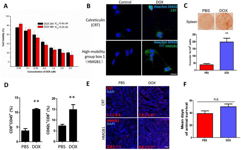
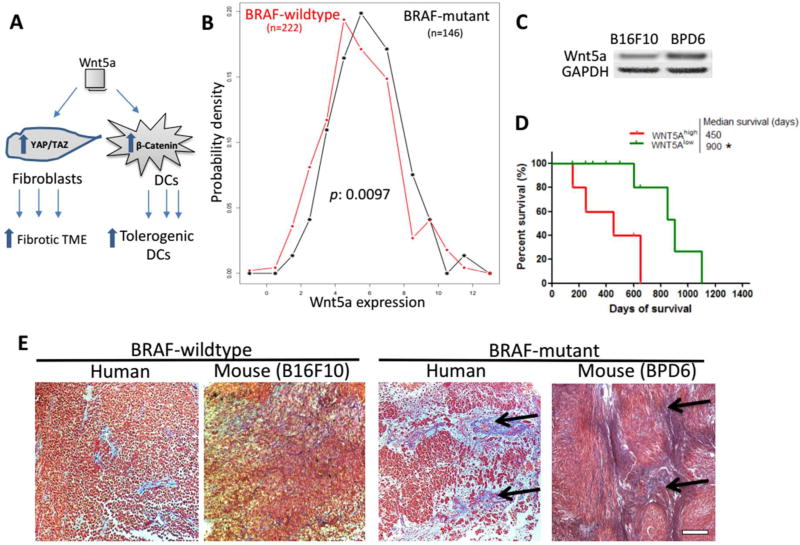
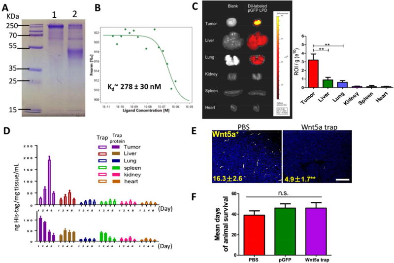
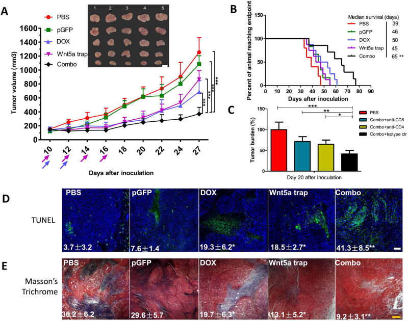
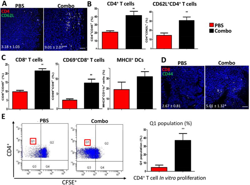
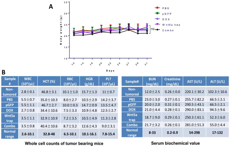
Development of an effective treatment against advanced tumors remains a major challenge for cancer immunotherapy. Approximately 50% of human melanoma is driven by B-Raf proto-oncogene mutation (BRAF mutant). Tumors with such mutation are desmoplastic, highly immunosuppressive, and often resistant to immune checkpoint therapies. We have shown that immunotherapy mediated by low-dose doxorubicin-induced immunogenic cell death was only partially effective for this type of tumor and not effective in long-term inhibition of tumor progression. Wnt family member 5A (Wnt5a), a signaling protein highly produced by BRAF mutant melanoma cells, has been implicated in inducing dendritic cell tolerance and tumor fibrosis, thus hindering effective antigen presentation and T-cell infiltration. We hypothesized that Wnt5a is a key molecule controlling the immunosuppressive tumor microenvironment in metastatic melanoma. Accordingly, we have designed and generated a trimeric trap protein, containing the extracellular domain of Fizzled 7 receptor that binds Wnt5a with a Kd ∼ 278 nM. Plasmid DNA encoding for the Wnt5a trap was delivered to the tumor by using cationic lipid-protamine-DNA nanoparticles. Expression of Wnt5a trap in the tumor, although transient, was greater than that of any other major organs including liver, resulting in a significant reduction of the Wnt5a level in the tumor microenvironment without systematic toxicity. Significantly, combination of Wnt5a trapping and low-dose doxorubicin showed great tumor growth inhibition and host survival prolongation. Our findings indicated that efficient local Wnt5a trapping significantly remodeled the immunosuppressive tumor microenvironment to facilitate immunogenic cell-death-mediated immunotherapy.
Keywords: B-Raf proto-oncogene mutant melanoma; Wnt family member 5A; immune trap; immunogenic cell death; nanoparticle; tumor microenvironment.
Conflict of interest statement
Figures

Similar articles
-
WNT5A enhances resistance of melanoma cells to targeted BRAF inhibitors.J Clin Invest. 2014 Jul;124(7):2877-90. doi: 10.1172/JCI70156. Epub 2014 May 27. J Clin Invest. 2014. PMID: 24865425 Free PMC article.
-
Inhibition of Age-Related Therapy Resistance in Melanoma by Rosiglitazone-Mediated Induction of Klotho.Clin Cancer Res. 2017 Jun 15;23(12):3181-3190. doi: 10.1158/1078-0432.CCR-17-0201. Epub 2017 Feb 23. Clin Cancer Res. 2017. PMID: 28232477 Free PMC article.
-
Immune Responses to BRAF-Targeted Therapy in Melanoma: Is Targeted Therapy Immunotherapy?Crit Rev Oncog. 2016;21(1-2):83-91. doi: 10.1615/CritRevOncog.2016017150. Crit Rev Oncog. 2016. PMID: 27481005 Review.
-
Combination therapy targeting the elevated interleukin-6 level reduces invasive migration of BRAF inhibitor-resistant melanoma cells.Mol Oncol. 2019 Feb;13(2):480-494. doi: 10.1002/1878-0261.12433. Epub 2019 Jan 10. Mol Oncol. 2019. PMID: 30582770 Free PMC article.
-
Tumor microenvironment changes leading to resistance of immune checkpoint inhibitors in metastatic melanoma and strategies to overcome resistance.Pharmacol Res. 2017 Sep;123:95-102. doi: 10.1016/j.phrs.2017.07.006. Epub 2017 Jul 6. Pharmacol Res. 2017. PMID: 28690075 Review.
Cited by
-
HDAC9 deficiency promotes tumor progression by decreasing the CD8+ dendritic cell infiltration of the tumor microenvironment.J Immunother Cancer. 2020 Jun;8(1):e000529. doi: 10.1136/jitc-2020-000529. J Immunother Cancer. 2020. PMID: 32554611 Free PMC article.
-
Emerging Nano-/Microapproaches for Cancer Immunotherapy.Adv Sci (Weinh). 2019 Jan 13;6(6):1801847. doi: 10.1002/advs.201801847. eCollection 2019 Mar 20. Adv Sci (Weinh). 2019. PMID: 30937265 Free PMC article. Review.
-
Membrane-core nanoparticles for cancer nanomedicine.Adv Drug Deliv Rev. 2020;156:23-39. doi: 10.1016/j.addr.2020.05.005. Epub 2020 May 22. Adv Drug Deliv Rev. 2020. PMID: 32450105 Free PMC article. Review.
-
Opportunities, obstacles and challenges of nano-immunotherapy in melanoma.Front Immunol. 2025 Aug 8;16:1611423. doi: 10.3389/fimmu.2025.1611423. eCollection 2025. Front Immunol. 2025. PMID: 40861441 Free PMC article. Review.
-
Disharmonic Inflammatory Signatures in COVID-19: Augmented Neutrophils' but Impaired Monocytes' and Dendritic Cells' Responsiveness.Cells. 2020 Sep 29;9(10):2206. doi: 10.3390/cells9102206. Cells. 2020. PMID: 33003471 Free PMC article.
References
-
- Gloster HM, Jr, Brodland DG. The Epidemiology of Skin Cancer. Dermatol. Surg. 1996;22:217–226. - PubMed
-
- Davies H, Bignell GR, Cox C, Stephens P, Edkins S, Clegg S, Teague J, Woffendin H, Garnett MJ, Bottomley W, Davis N, Dicks E, Ewing R, Floyd Y, Gray K, Hall S, Hawes R, Hughes J, Kosmidou V, Menzies A, et al. Mutations of the Braf Gene in Human Cancer. Nature. 2002;417:949–954. - PubMed
-
- Crosby T, Fish R, Coles B, Mason MD. Systemic Treatments for Metastatic Cutaneous Melanoma. Cochrane Database Syst Rev. 2000:CD001215. - PubMed
-
- Liu Q, Das M, Liu Y, Huang L. Targeted Drug Delivery to Melanoma. Adv. Drug Deliv Rev. 2017 - PubMed
Publication types
MeSH terms
Substances
Grants and funding
LinkOut - more resources
Full Text Sources
Other Literature Sources
Medical
Research Materials
Miscellaneous
