Review
. 2018 Jan 22;10(1):7.
doi: 10.1186/s13195-017-0334-y.

Extended FTLD pedigree segregating a Belgian GRN-null mutation: neuropathological heterogeneity in one family

Affiliations
Review

Extended FTLD pedigree segregating a Belgian GRN-null mutation: neuropathological heterogeneity in one family

Anne Sieben et al. Alzheimers Res Ther. .

Abstract

Background: In this paper, we describe the clinical and neuropathological findings of nine members of the Belgian progranulin gene (GRN) founder family. In this family, the loss-of-function mutation IVS1 + 5G > C was identified in 2006. In 2007, a clinical description of the mutation carriers was published that revealed the clinical heterogeneity among IVS1 + 5G > C carriers. We report our comparison of our data with the published clinical and neuropathological characteristics of other GRN mutations as well as other frontotemporal lobar degeneration (FTLD) syndromes, and we present a review of the literature.

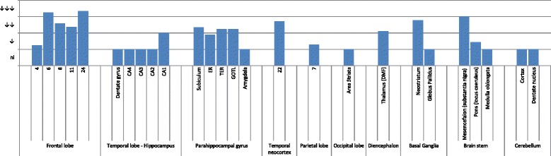
Methods: For each case, standardized sampling and staining were performed to identify proteinopathies, cerebrovascular disease, and hippocampal sclerosis.

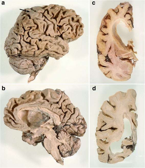
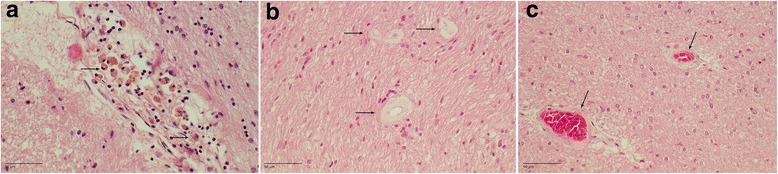
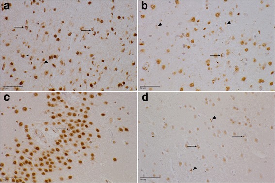
Results: The neuropathological substrate in the studied family was compatible in all cases with transactive response DNA-binding protein (TDP) proteinopathy type A, as expected. Additionally, most of the cases presented also with primary age-related tauopathy (PART) or mild Alzheimer's disease (AD) neuropathological changes, and one case had extensive Lewy body pathology. An additional finding was the presence of cerebral small vessel changes in every patient in this family.

Conclusions: Our data show not only that the IVS1 + 5G > C mutation has an exclusive association with FTLD-TDP type A proteinopathy but also that other proteinopathies can occur and should be looked for. Because the penetrance rate of the clinical phenotype of carriers of GRN mutations is age-dependent, further research is required to investigate the role of co-occurring age-related pathologies such as AD, PART, and cerebral small vessel disease.

Keywords: Cerebral small vessel disease (SVD); FTD; FTD-GRN; FTLD; FTLD-TDP; Frontotemporal dementia; Frontotemporal lobar degeneration.


Conflict of interest statement

Consent for publication

The patients and/or their legal representatives gave their consent for publication.

Competing interests

The authors declare that they have no competing interests.

Publisher’s Note

Springer Nature remains neutral with regard to jurisdictional claims in published maps and institutional affiliations.

Figures

Fig. 1
Fig. 1
Macroscopic images. a Lateral view of the right hemisphere in DR2.3. Note the frontal and temporal, and to a lesser extent parietal, atrophy. Area 4 (arrow) is relatively preserved. b Medial view of right hemisphere in DR2.3. The superior frontal gyrus is atrophied, whereas the straight gyrus is relatively spared. c Coronal section of the right hemisphere trough the head of the caudate nucleus in DR2.3. There is a flattened, nearly concave aspect of the caudate nucleus. d Coronal sections trough the parietotemporal lobes in DR8.1. The lateral ventricle is dilated, especially the temporal horn. Temporal atrophy is more pronounced than parietal atrophy
Fig. 2
Fig. 2
Grouped histopathological findings. Entorhinal cortex (ER), transentorhinal cortex (TER), gyrus occipitotemporalis lateralis (GOTL), dorsomedial formation (DMF), and status pigmentatus (SP). Semiquantitative analysis of the average neuronal cell loss in every region of interest. nl Normal, ↓ Slight neuronal loss, ↓↓ Moderate neuronal loss, ↓↓↓ Severe neuronal loss
Fig. 3
Fig. 3
Cerebral small vessel disease. a DR2.3. H&E stain of perivascular hemosiderin deposits in the hippocampus. Arrows show many siderophages. b DR25.5. H&E stain of the corpus callosum. Arrows show thickening of the arteriolar wall. c DR25.5. H&E stain of the temporal white matter. Arrows show thickening of the arteriolar walls with mild perivascular demyelination
Fig. 4
Fig. 4
Transactive response DNA-binding protein (TDP) pathology (paraffin-embedded sections stained with antihyperphosphorylated TDP-43 antibody). a DR25.5 area 6. There is a moderate amount of neuronal intracytoplasmic inclusions (NCIs) (arrows), mainly in the second cortical layer. The dystrophic neurites (arrowhead) are more evenly spread throughout the entire cortex. b DR2.3 superior temporal gyrus. Mild to moderate TDP-43 proteinopathy type A with NCIs (arrow) and dystrophic neurites (arrowheads) can be seen. c DR28.1 dentate gyrus. There are scarce NCIs (arrow) in the granular layer. d DR31.1 area striata. A mild amount of NCIs (arrows) and dystrophic neurites (arrowheads) in the second cortical layer can be seen
Fig. 5
Fig. 5
Transactive response DNA-binding protein pathology in different brain areas. ± (sparse (0–1/mm2)), + (mild (< 5/mm2)), ++ (moderate (5–20/mm2)), +++ (extensive (> 20/mm)). The average lesion load is shown for the most important regions of interest. Striped bar represents the amount of neuronal intracytoplasmic inclusions, whereas the dark bar represents the amount of short dystrophic neurites. With the dotted bar, the neuronal intranuclear inclusions are shown. NCI Neuronal intracytoplasmic inclusions, DN Dystrophic neurites, NII Nuclear intraneuronal inclusions, DMF Dorsomedial formation, ER Entorhinal cortex, TER Transentorhinal cortex, GOTL Gyrus occipitotemporalis lateralis, DG Dentate gyrus

References

    1. Rascovsky K, et al. Sensitivity of revised diagnostic criteria for the behavioural variant of frontotemporal dementia. Brain. 2011;134(Pt 9):2456–77. doi: 10.1093/brain/awr179. - DOI - PMC - PubMed
    1. Neary D, et al. Frontotemporal lobar degeneration: a consensus on clinical diagnostic criteria. Neurology. 1998;51(6):1546–54. doi: 10.1212/WNL.51.6.1546. - DOI - PubMed
    1. Gorno-Tempini ML, et al. Classification of primary progressive aphasia and its variants. Neurology. 2011;76(11):1006–14. doi: 10.1212/WNL.0b013e31821103e6. - DOI - PMC - PubMed
    1. Hutton M, et al. Association of missense and 5′-splice-site mutations in tau with the inherited dementia FTDP-17. Nature. 1998;393(6686):702–5. doi: 10.1038/31508. - DOI - PubMed
    1. Poorkaj P, et al. Tau is a candidate gene for chromosome 17 frontotemporal dementia. Ann Neurol. 1998;43(6):815–25. doi: 10.1002/ana.410430617. - DOI - PubMed

Publication types

LinkOut - more resources