. 2018 Jan 9;10(1):1.
doi: 10.1186/s13195-017-0329-8.

Diffusion kurtosis imaging allows the early detection and longitudinal follow-up of amyloid-β-induced pathology

Affiliations

Diffusion kurtosis imaging allows the early detection and longitudinal follow-up of amyloid-β-induced pathology

Jelle Praet et al. Alzheimers Res Ther. .

Abstract

Background: Alzheimer's disease (AD) is a progressive neurodegenerative disorder and the most common cause of dementia in the elderly population. In this study, we used the APP/PS1 transgenic mouse model to explore the feasibility of using diffusion kurtosis imaging (DKI) as a tool for the early detection of microstructural changes in the brain due to amyloid-β (Aβ) plaque deposition.

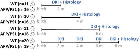
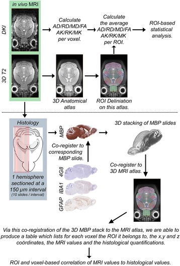
Methods: We longitudinally acquired DKI data of wild-type (WT) and APP/PS1 mice at 2, 4, 6 and 8 months of age, after which these mice were sacrificed for histological examination. Three additional cohorts of mice were also included at 2, 4 and 6 months of age to allow voxel-based co-registration between diffusion tensor and diffusion kurtosis metrics and immunohistochemistry.

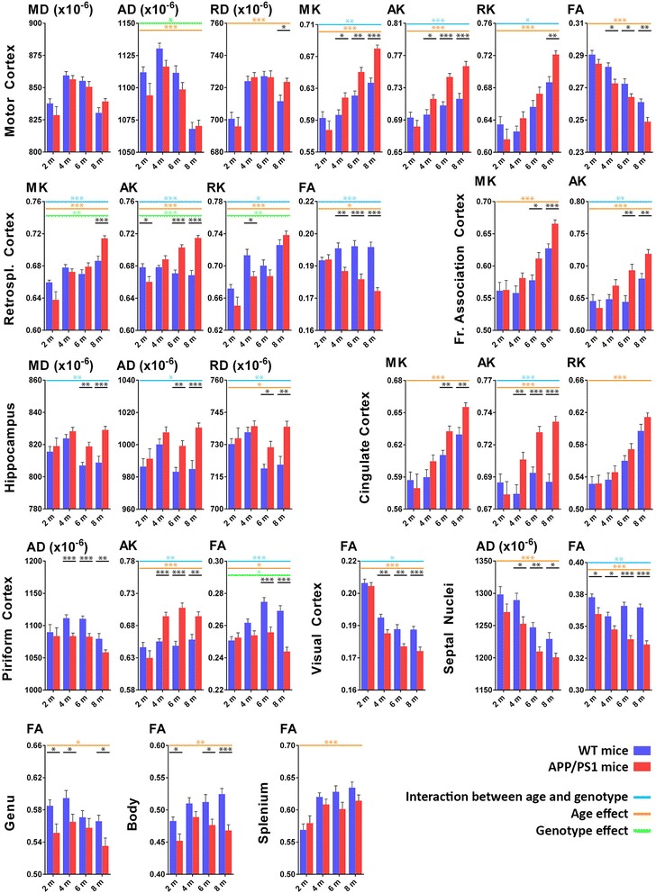
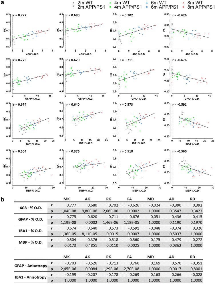
Results: Changes were observed in diffusion tensor (DT) and diffusion kurtosis (DK) metrics in many of the 23 regions of interest that were analysed. Mean and axial kurtosis were greatly increased owing to Aβ-induced pathological changes in the motor cortex of APP/PS1 mice at 4, 6 and 8 months of age. Additionally, fractional anisotropy (FA) was decreased in APP/PS1 mice at these respective ages. Linear discriminant analysis of the motor cortex data indicated that combining diffusion tensor and diffusion kurtosis metrics permits improved separation of WT from APP/PS1 mice compared with either diffusion tensor or diffusion kurtosis metrics alone. We observed that mean kurtosis and FA are the critical metrics for a correct genotype classification. Furthermore, using a newly developed platform to co-register the in vivo diffusion-weighted magnetic resonance imaging with multiple 3D histological stacks, we found high correlations between DK metrics and anti-Aβ (clone 4G8) antibody, glial fibrillary acidic protein, ionised calcium-binding adapter molecule 1 and myelin basic protein immunohistochemistry. Finally, we observed reduced FA in the septal nuclei of APP/PS1 mice at all ages investigated. The latter was at least partially also observed by voxel-based statistical parametric mapping, which showed significantly reduced FA in the septal nuclei, as well as in the corpus callosum, of 8-month-old APP/PS1 mice compared with WT mice.

Conclusions: Our results indicate that DKI metrics hold tremendous potential for the early detection and longitudinal follow-up of Aβ-induced pathology.

Keywords: APP/PS1; Alzheimer’s disease; Diffusion kurtosis imaging; Diffusion tensor imaging; Magnetic resonance imaging.


Conflict of interest statement

Authors’ information

Not applicable.

Ethics approval and consent to participate

All experimental procedures were performed in accordance with European guidelines (2010/63/EU) and were approved by the University of Antwerp Ethics Committee for Animal Experiments (approval number 2012-46).

Consent for publication

Not applicable.

Competing interests

The authors declare that they have no competing interests.

Publisher’s Note

Springer Nature remains neutral with regard to jurisdictional claims in published maps and institutional affiliations.

Figures

Fig. 1
Fig. 1
Experimental setup of the study. Diffusion kurtosis imaging (DKI) was performed in three cohorts of male wild-type (WT) and APP/PS1 mice at 2, 4 and 6 months of age, and these mice were sacrificed for histological analysis thereafter. In a fourth, longitudinal cohort of male WT and APP/PS1 mice, we performed DKI at 2, 4, 6 and 8 months of age and thereafter killed these mice for histological analysis
Fig. 2
Fig. 2
Schematic overview of the data acquisition and analysis pipeline used in this study. AD Axial diffusivity, AK Axial kurtosis, DKI Diffusion kurtosis imaging, FA Fractional anisotropy, 4G8 Anti-amyloid-β (clone 4G8) antibody, GFAP Glial fibrillary acidic protein, IBA1 Ionised calcium-binding adapter molecule 1, MBP Myelin basic protein, MD Mean diffusivity, MK Mean kurtosis, MRI Magnetic resonance imaging, RD Radial diffusivity, RK Radial kurtosis, ROI Region of interest
Fig. 3
Fig. 3
Region of interest (ROI)-based DKI analysis. The most interesting differences in diffusion tensor and diffusion kurtosis metrics of several grey and white matter ROIs. We show only data from the longitudinal cohort of wild-type WT (blue bars) and APP/PS1 mice (red bars) at 2, 4, 6 and 8 months of age. Shown are the mean diffusivity (MD), axial diffusivity (AD) and radial diffusivity (RD); the mean kurtosis (MK), axial kurtosis (AK) and radial kurtosis (RK); and the fractional anisotropy (FA). Significant differences between WT and APP/PS1 mice at any given time point are shown in black; genotype effect is indicated by green lines; age effect is indicated by orange lines; and interaction between age and genotype is indicated by blue lines. *p < 0.05, **p < 0.01 and ***p < 0.001
Fig. 4
Fig. 4
Linear discriminant analysis and least absolute shrinkage and selection operator (LASSO) analysis of the motor cortex. a The frequency distribution histograms of the misclassification error (MCE) of the motor cortex are shown (percentage of animals which were attributed the wrong genotype). The MCE is calculated at 2, 4, 6 and 8 months of age when using only the diffusion tensor (DT) metrics (top row), the diffusion kurtosis (DK) metrics (middle row) or a combination of the DT and DK metrics (bottom row). The red lines indicate the average MCE. b LASSO analysis was done to determine which of the DTI or DKI metrics contribute the most to a correct classification of the genotype. The heat map shows all 100 iterations of the LASSO analysis for all metrics, where green indicates that the metric was used and red indicates that the metric was not used. On the right, we show the percentage of times that the metric contributed to a correct genotype classification in all of these 100 iterations (percent prevalence)
Fig. 5
Fig. 5
Representative histological images of the motor cortex of the wild-type (WT) and APP/PS1 mice (left and right columns, respectively) at 2, 4, 6 and 8 months of age (top to bottom rows). The different panels show the anti-amyloid-β (clone 4G8) antibody staining for amyloid-β plaques (a), the myelin basic protein staining for myelin basic protein (b), the glial fibrillary acidic protein staining for astrogliosis (c) and the ionised calcium-binding adapter molecule 1staining for microgliosis (d)
Fig. 6
Fig. 6
Region of interest-based histological correlation analysis. a Graphs showing the Pearson correlations between mean kurtosis (MK), axial kurtosis (AK), radial kurtosis (RK) and fractional anisotropy (FA) and percent optical density (%O.D.) of anti-amyloid-β (clone 4G8) antibody (4G8), glial fibrillary acidic protein (GFAP), ionised calcium-binding adapter molecule 1 (IBA1) and myelin basic protein (MBP). The graph includes data from the wild-type (WT) mice (triangles) and APP/PS1 mice (circles) at 2 months of age (grey), 4 months of age (green), 6 months of age (blue) and 8 months of age (red). b Pearson correlation values (r) and p values of these correlations between the different diffusion tensor and diffusion kurtosis metrics and the histological parameters
Fig. 7
Fig. 7
Voxel-based statistical parametric mapping. a Statistical parametric maps at two different levels in the brains of mice at 8 months of age. On these images, for each of the seven metrics (mean diffusivity [MD], axial diffusivity [AD], radial diffusivity [RD], mean kurtosis [MK], axial kurtosis [AK], radial kurtosis [RK] and fractional anisotropy [FA]), the voxels with a decreased value in APP/PS1 mice compared with wild-type (WT) mice are shown in blue, and the voxels with an increased value in APP/PS1 mice compared with WT mice are shown in red. Only voxels with a false discovery rate-corrected p > 0.05 are shown. b Close-up of the FA statistical parametric map showing reduced FA in APP/PS1 mice in the septal nuclei at 8 months of age (blue voxels). c Voxel-based correlation values between the percent optical density (%O.D.) anti-amyloid-β (clone 4G8) antibody (4G8), myelin basic protein (MBP), glial fibrillary acidic protein (GFAP) and ionised calcium-binding adapter molecule 1 (IBA1) and the diffusion tensor metrics (DT), the diffusion kurtosis metrics (DK), and the DT and DK metrics (DT/DK)

References

    1. Alzheimer’s Association 2015 Alzheimer’s disease facts and figures. Alzheimers Dement. 2015;11:332–384. doi: 10.1016/j.jalz.2015.02.003. - DOI - PubMed
    1. Howard R, McShane R, Lindesay J, Ritchie C, Baldwin A, Barber R, et al. Donepezil and memantine for moderate-to-severe Alzheimer’s disease. N Engl J Med. 2012;366:893–903. doi: 10.1056/NEJMoa1106668. - DOI - PubMed
    1. Leifer BP. Early diagnosis of Alzheimer’s disease: clinical and economic benefits. J Am Geriatr Soc. 2003;51(5 Suppl Dementia):S281–S288. doi: 10.1046/j.1532-5415.5153.x. - DOI - PubMed
    1. Liu-Seifert H, Siemers E, Holdridge KC, Andersen SW, Lipkovich I, Carlson C, et al. Delayed-start analysis: mild Alzheimer’s disease patients in solanezumab trials, 3.5 years. Alzheimers Dement (N Y) 2015;1:111–121. - PMC - PubMed
    1. McKhann G, Knopman DS, Chertkow H, Hymann B, Jack CR, Jr, Kawas C, et al. The diagnosis of dementia due to Alzheimer’s disease: recommendations from the National Institute on Aging-Alzheimer’s Association workgroups on diagnostic guidelines for Alzheimer’s disease. Alzheimers Dement. 2011;7:263–269. doi: 10.1016/j.jalz.2011.03.005. - DOI - PMC - PubMed

Publication types