. 2018 Jan 25;51(1):1701214.
doi: 10.1183/13993003.01214-2017. Print 2018 Jan.

The inflammatory cell landscape in the lungs of patients with idiopathic pulmonary arterial hypertension

Affiliations

The inflammatory cell landscape in the lungs of patients with idiopathic pulmonary arterial hypertension

Leigh M Marsh et al. Eur Respir J. .

Abstract

Increasing evidence points towards an inflammatory component underlying pulmonary hypertension. However, the conclusive characterisation of multiple inflammatory cell populations in the lung is challenging due to the complexity of marker specificity and tissue inaccessibility. We used an unbiased computational flow cytometry approach to delineate the inflammatory landscape of idiopathic pulmonary arterial hypertension (IPAH) and healthy donor lungs.Donor and IPAH samples were discriminated clearly using principal component analysis to reduce the multidimensional data obtained from single-cell flow cytometry analysis. In IPAH lungs, the predominant CD45+ cell type switched from neutrophils to CD3+ T-cells, with increases in CD4+, CD8+ and γδT-cell subsets. Additionally, diversely activated classical myeloid-derived dendritic cells (CD14-HLA-DR+CD11c+CD1a+/-) and nonclassical plasmacytoid dendritic cells (pDCs; CD14-CD11c-CD123+HLA-DR+), together with mast cells and basophils, were more abundant in IPAH samples. We describe, for the first time, the presence and regulation of two cell types in IPAH, γδT-cells and pDCs, which link innate and adaptive immunity.With our high-throughput flow cytometry with multidimensional dataset analysis, we have revealed the interactive interplay between multiple inflammatory cells is a crucial part of their integrative network. The identification of γδT-cells and pDCs in this disease potentially provides a missing link between IPAH, autoimmunity and inflammation.


Conflict of interest statement

Conflict of interest: Disclosures can be found alongside this article at erj.ersjournals.com

Figures

FIGURE 1
FIGURE 1
Schematic representation of analysed cell populations identified during flow cytometric analysis. Hierarchal organisation of all haematopoietic derived CD45+ cells of both lymphoid and myeloid lineages. NK: natural killer cells; NKT: natural killer T-cells; PMN: polymorphonuclear neutrophils; Baso: basophils; Eos: eosinophils; Mono: monocytes; Int: intermediate monocytes; Nonclass: nonclassical monocytes; DC: dendritic cells; pDC: plasmacytoid DC; Macs: macrophages. Italic letters (a–p) represent common cell populations, as identified in figure 2.
FIGURE 2
FIGURE 2
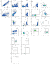
Overview of gating strategy used in flow cytometric analysis. Analysed populations were first gated to remove cell clumps and debris, and dead cells were removed by viability exclusion. All leukocytes were selected using CD45-positivity and then divided into separate cell populations as shown. FSC-H: forward scatter height; FSC-A: forward scatter area; SSC: side scatter; HLA-DR: human leukocyte antigen – antigen D related; pDCs: plasmacytoid dendritic cells; NK: natural killer cells; NKT: natural killer T-cells. Boxes and italic lettering (a–p) represent common cell populations, as identified in figure 1.
FIGURE 3
FIGURE 3
Principal component analysis plots of flow cytometric data separates idiopathic pulmonary arterial hypertension (IPAH) from control samples. The log odds ratios of subset analysis data obtained by flow cytometry using manual gating were used to construct a similarity matrix for the principal component analysis. The first two principal components (PC) with respective percentage variation are depicted. Bivariate plots of dimensionality reduction from a) 12 lungs (seven donor/five IPAH) including 21 cell populations; b) 31 lungs (15 donor/16 IPAH) and 18 cell populations.
FIGURE 4
FIGURE 4
Global changes in inflammatory profiles. a) Sunburst plots showing average distribution of all cell populations in donor lungs and lungs from patients with idiopathic pulmonary arterial hypertension; b) heatmap representation presenting the patient-to-patient variability of the log odds ratio derived from cell number per mg tissue data for 18 individual cell populations. CTL: cytotoxic CD8+ T-lymphocytes; PMN: polymorphonuclear neutrophils; Macs: macrophages; Monos: monocytes; Int: intermediate; Nonclass: nonclassical; DCs: dendritic cells; pDCs: plasmacytoid DCs.
FIGURE 5
FIGURE 5
T-stochastic neighbour embedding (t-SNE) visualisation and regulated cell populations in lung samples. a) t-SNE composite dimension plots of down-sampled and concatenated CD45+ cells derived from flow cytometric data with overlaid manually gated cell populations. b) Regulated cell populations differentially abundant between idiopathic pulmonary arterial hypertension and donor lungs as identified by flow cytometry. DCs: dendritic cells; pDCs: plasmacytoid dendritic cells. Boxplots show median and interquartile range.
FIGURE 6
FIGURE 6
Abundance of cell populations in isolated pulmonary arteries. The presence of multiple cell populations (as shown for lung homogenate samples) measured in third- and fourth-generation pulmonary arteries by flow cytometry. a) T-cells; b) CD4+ T-cells; c) CD8+ T-cells; d) γδT-cells; e) mast cells; f) basophils; g) dendritic cells (CD209- CD11c+); h) dendritic cells (CD209- CD11c+ CD1a+); i) plasmacytoid dendritic cells. IPAH: idiopathic pulmonary arterial hypertension. Boxplots show median and interquartile range.
FIGURE 7
FIGURE 7
Presence and localisation of plasmacytoid dendritic cells (pDCs) and γδT-cells in the lung. Immunofluorescence staining and quantification of idiopathic pulmonary arterial hypertension (IPAH) and donor lung tissue against a,b) CD123 (red) and CD304 (green) to detect pDCs (CD123+CD304+) and c,d) TCRγ (green) to detect γδT-cells. Smooth muscle actin (SMA) staining is shown in purple; nuclear staining via DAPI (blue). Smaller panels represent magnified area as shown in larger panels. White arrows indicate cells of interest, red and green arrows indicate single positive cells. Neg control: staining performed in the absence of primary antibodies. Scale bars=20 µm.
FIGURE 8
FIGURE 8
Idiopathic pulmonary arterial hypertension (IPAH) patients do not possess elevated plasmacytoid dendritic cell (pDC) numbers in the peripheral blood. a) Gating strategy for the flow cytometric quantification of circulating pDCs (CD14CD11cCD123+CD304+HLR-DR+) and basophils (CD14CD11cCD123+CD304FceRi+) in peripheral blood. b) Scatterplot and boxplot overlays of quantified total CD11cCD123+ cells, pDCs and basophils. Boxplots show median and interquartile range.

Comment in

References

    1. Stacher E, Graham BB, Hunt JM, et al. Modern age pathology of pulmonary arterial hypertension. Am J Respir Crit Care Med 2012; 186: 261–272. - PMC - PubMed
    1. Nicolls MR, Voelkel NF. The roles of immunity in the prevention and evolution of pulmonary arterial hypertension. Am J Respir Crit Care Med 2017; 195: 1292–1299. - PMC - PubMed
    1. Heath D. Pulmonary hypertension in pulmonary parenchymal disease. Cardiovasc Clin 1972; 4: 79–96. - PubMed
    1. Tuder RM, Groves B, Badesch DB, et al. Exuberant endothelial cell growth and elements of inflammation are present in plexiform lesions of pulmonary hypertension. Am J Pathol 1994; 144: 275–285. - PMC - PubMed
    1. Perros F, Dorfmüller P, Souza R, et al. Dendritic cell recruitment in lesions of human and experimental pulmonary hypertension. Eur Respir J 2007; 29: 462–468. - PubMed

Publication types

MeSH terms

Substances