Review
. 2018 Jan 25;9(2):115.
doi: 10.1038/s41419-017-0061-0.

Defining the role of the tumor vasculature in antitumor immunity and immunotherapy

Affiliations
Review

Defining the role of the tumor vasculature in antitumor immunity and immunotherapy

Marco B Schaaf et al. Cell Death Dis. .

Abstract

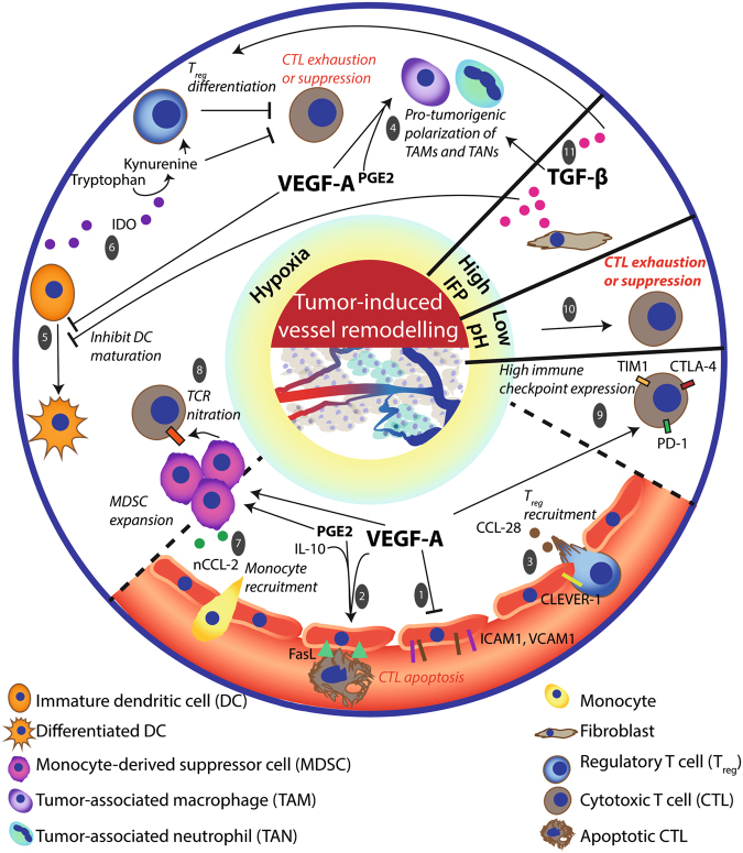
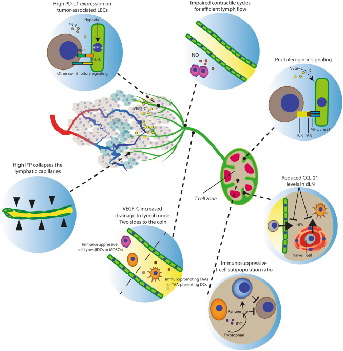
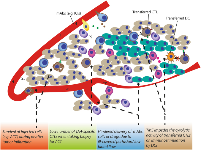
It is now well established that cancer cells co-exist within a complex environment with stromal cells and depend for their growth and dissemination on tight and plastic interactions with components of the tumor microenvironment (TME). Cancer cells incite the formation of new blood and lymphatic vessels from preexisting vessels to cope with their high nutrient/oxygen demand and favor tumor outgrowth. Research over the past decades has highlighted the crucial role played by tumor-associated blood and lymphatic vasculature in supporting immunoevasion and in subverting T-cell-mediated immunosurveillance, which are the main hallmarks of cancers. The structurally and functionally aberrant tumor vasculature contributes to the protumorigenic and immunosuppressive TME by maintaining a cancer cell's permissive environment characterized by hypoxia, acidosis, and high interstitial pressure, while simultaneously generating a physical barrier to T cells' infiltration. Recent research moreover has shown that blood endothelial cells forming the tumor vessels can actively suppress the recruitment, adhesion, and activity of T cells. Likewise, during tumorigenesis the lymphatic vasculature undergoes dramatic remodeling that facilitates metastatic spreading of cancer cells and immunosuppression. Beyond carcinogenesis, the erratic tumor vasculature has been recently implicated in mechanisms of therapy resistance, including those limiting the efficacy of clinically approved immunotherapies, such as immune checkpoint blockers and adoptive T-cell transfer. In this review, we discuss emerging evidence highlighting the major role played by tumor-associated blood and lymphatic vasculature in thwarting immunosurveillance mechanisms and antitumor immunity. Moreover, we also discuss novel therapeutic approaches targeting the tumor vasculature and their potential to help overcoming immunotherapy resistance.


Conflict of interest statement

The authors declare that they have no competing financial interests.

Figures

Fig. 1
Fig. 1. Tumor-associated blood vasculature is a major influencer of the tumor microenvironment (TME)
(Upper left panel) A well-organized vessel network ensures full-covering of nutrient supply. (Lower left panel) These vessels are matured with an endothelial cell layer surrounded by a basement membrane and pericytes (like smooth muscle cells). The endothelial layer is characterized by tight intercellular junctions. Oppositely, due to high pro-angiogenic signaling, the network of tumor-associated blood vessels (upper right panel) is chaotic, low in pericyte coverage and has loose inter-endothelial cell junctions (lower right panel). This generates leaky vessels that increases interstitial fluid (IFP) pressure. Common blunt-ended or collapsed vessels results in tumor regions that are starved from nutrients including oxygen (hypoxic cells indicated in green). Moreover, the glycolytic nature of the (hypoxic) tumor cell acidifies the pH in the TME
Fig. 2
Fig. 2. The nature of the TME influences immune cell composition and hampers antitumor immunity
First, hypoxia is a common feature of the TME caused by the abnormal vascular structure and function. Dysregulated adhesion [1] and differential admittance among immune cell types is caused by several hypoxia-related factors in the TME, including VEGF-A, PGE2 and IL-10. Together these induce FasL expression on ECs that affects survival of effector T cells (rather than Tregs). [2] In addition, expression of CLEVER-1/stabilin-1 on tumor-ECs and hypoxia-related chemokine CCL-28 in the TME further aid the recruitment of, preferentially, Tregs. [3] The hypoxic TME recruits monocytes that give rise to MDSC, TAM, and TAN populations [4] in the tumor and induces a differential and functional immature phenotype of DCs. [5] This collectively supports an immunosuppressive TME. Immature DCs produce IDO to favor Treg differentiation from naive T cells and inhibit CTL function. [6] MDSCs are a source of reactive nitrogen species that nitrate CCL-2 and tyrosines of the T-cell receptor that recruits more monocytes [7] and impedes CTL antigen recognition, [8] respectively. Moreover, VEGF-A induces the expression of PD-1, TIM-3, and CTLA-4 on CTLs to render them more susceptible to functional inhibition. [9] Second, as a result of a more glycolytic metabolism the TME acidifies (low pH), thereby inhibiting the induction of antigen specific CTLs. [10] Third, the leaky tumor vessels induce a high interstitial fluid pressure (IFP) that leads to high TGF-β production that is also implicated in TAM M2 polarization, maintaining immature DC phenotype and differentiation and proliferation of Tregs. [11]
Fig. 3
Fig. 3. The effects of tumor-associated lymphatic endothelium on antitumor immunity
The lymphatic vessels (green) guide antigens and DCs to lymph nodes to facilitate the DC–T cell interaction to prime T cells (only if the LN microenvironment allows this to be productive). Notably, lymphatic vessels are more common peritumorally, while intratumoral vessels are prone to collapse. Moreover, defects in contractile events for lymph flow impair drainage. Thus tumor drainage, albeit physically hampered in a tumor, is required for developing antitumor immunity. Importantly, additional LEC features (intrinsic or tumor induced) counteract the induction of an adaptive immune response. This is exemplified by the increased PD-L1 expression and protolerogenic cell surface protein composition (co-inhibitory over co-stimulatory factors). Drainage of immunosuppressive immune cell types (e.g., MDSCs, immature DCs) influence the LN microenvironment to favor immunosuppressive populations (e.g., Treg and MDSCs) that facilitate lymphovascular premetastatic niche formation. Moreover, reduced CCL-21 levels in dLNs diminish the opportunity for DCs and naive T cells to interact and impairs T-cell retention for efficient expansion before LN egress.
Fig. 4
Fig. 4. Hurdles established by the tumor vasculature that limit immunotherapy efficacy
As discussed above, the TME often thwart CTL presence in the TME due to inducing apoptosis/ill-adhesion or by functional inhibition even when infiltrated. This low number of TAA-specific CTLs affects the harvest from tumor biopsies for adoptive T-cell transfer-based immunotherapy. Moreover, delivery of administered regimens including monoclonal antibodies, DCs, and T cells can be hindered due to ill-perfusion. Yet, the TME can still functionally inhibit the transferred DCs when infiltrated

References

    1. Hanahan D, Coussens LM. Accessories to the crime: functions of cells recruited to the tumor microenvironment. Cancer Cell. 2012;21:309–322. doi: 10.1016/j.ccr.2012.02.022. - DOI - PubMed
    1. Dunn GP, Old LJ, Schreiber RD. The immunobiology of cancer immunosurveillance and immunoediting. Immunity. 2004;21:137–148. doi: 10.1016/j.immuni.2004.07.017. - DOI - PubMed
    1. Vesely MD, Kershaw MH, Schreiber RD, Smyth MJ. Natural innate and adaptive immunity to cancer. Annu. Rev. Immunol. 2011;29:235–271. doi: 10.1146/annurev-immunol-031210-101324. - DOI - PubMed
    1. Blankenstein T, Coulie PG, Gilboa E, Jaffee EM. The determinants of tumour immunogenicity. Nat. Rev. Cancer. 2012;12:307–313. doi: 10.1038/nrc3246. - DOI - PMC - PubMed
    1. Weis SM, Cheresh DA. Tumor angiogenesis: molecular pathways and therapeutic targets. Nat. Med. 2011;17:1359–1370. doi: 10.1038/nm.2537. - DOI - PubMed

Publication types