. 2018 Jan 25;8(1):1603.
doi: 10.1038/s41598-017-16703-9.

Characterization of Nanodiamond-based anti-HIV drug Delivery to the Brain

Affiliations

Characterization of Nanodiamond-based anti-HIV drug Delivery to the Brain

Upal Roy et al. Sci Rep. .

Abstract

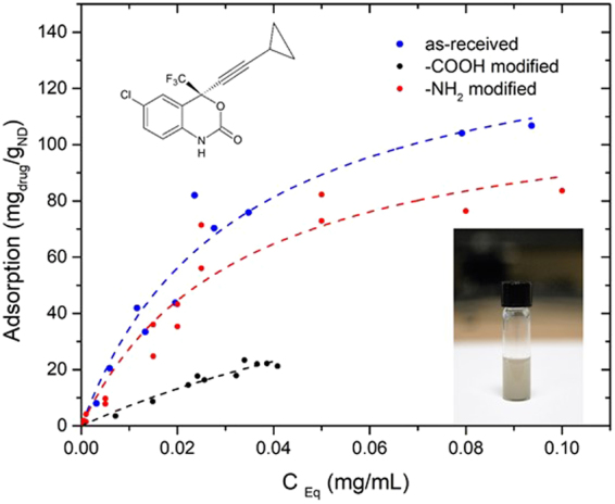
Human Immunodeficiency Virus Type 1 (HIV-1) remains one of the leading causes of death worldwide. Present combination antiretroviral therapy has substantially improved HIV-1 related pathology. However, delivery of therapeutic agents to the HIV reservoir organ like Central nervous system (CNS) remains a major challenge primarily due to the ineffective transmigration of drugs through Blood Brain Barrier (BBB). The recent advent of nanomedicine-based drug delivery has stimulated the development of innovative systems for drug delivery. In this regard, particular focus has been given to nanodiamond due to its natural biocompatibility and non-toxic nature-making it a more efficient drug carrier than other carbon-based materials. Considering its potential and importance, we have characterized unmodified and surface-modified (-COOH and -NH2) nanodiamond for its capacity to load the anti-HIV-1 drug efavirenz and cytotoxicity, in vitro. Overall, our study has established that unmodified nanodiamond conjugated drug formulation has significantly higher drug loading capacity than surface-modified nanodiamond with minimum toxicity. Further, this nanodrug formulation was characterized by its drug dissolution profile, transmigration through the BBB, and its therapeutic efficacy. The present biological characterizations provide a foundation for further study of in-vivo pharmacokinetics and pharmacodynamics of nanodiamond-based anti-HIV drugs.


Conflict of interest statement

A US patent has been issued on this work (9616022B1).

Figures

Figure 1
Figure 1
Characterization of ND: TEM images of (A) as-received ND, (B) –COOH and (C) –NH2 modified nanodiamonds. (D) Powder X-ray diffraction patterns of as-received ND in comparison with –COOH and –NH2 functional groups modified ND powders. The reflecting atomic planes of the diamond structure are denoted by Miller indices. (E) Raman spectra of (a) as-received ND, (b) –COOH and (c) –NH2 modified nanodiamonds.
Figure 2
Figure 2
Effect of ND, ND-COOH, & ND-NH2 on ROS production on SK-N-MC cells: Three formulations were exposed at different concentrations (10–1000 µg/ml) to SK-N-MC cells for 24 h. At the end of incubation, ROS production was measured in treated cells compared to untreated cells. The ROS production was measured in terms of mean ± SE relative fluorescence units (RFU) of eight independent experimental values. The statistical significance between untreated (ND) and ND-COOH treated cells were expressed as p values (**p < 0.001, ***p < 0.0003). There was no statistical significance between untreated (ND) and ND-NH2 groups.
Figure 3
Figure 3
Cytotoxicity of ND, ND-COOH, and ND-NH2 on SK-N-MC cells. The cells were treated with a range of concentrations (0.5–1000 µg/ml) for 24 h, separately. After incubation, MTS assay was performed and optical density (OD) was measured at 490nm. Graphical representation was made in terms of % survival of cells at different concentrations of three formulations corresponding to their OD values. Untreated cells (control) were considered as 100% viability and % survival was monitored based on control. The statistical significance between ND, ND-COOH, & ND-NH2 groups compared to control was expressed as p values (***p < 0.0001).
Figure 4
Figure 4
Adsorption isotherm of EFV on ND. The dotted line showed EFV adsorption by ND (blue), ND-NH2 (red) and ND-COOH (black), respectively. The adsorption values were calculated and represented as experimental points by Langmuir model. Inserts showed structural formula of efavirenz drug only and ND suspension in PBS buffer solution, respectively.
Figure 5
Figure 5
Drug dissolution study of nanodrug (ND) vs. Free drug (FD) in vitro. The released drug outside of dialysis bag was measured from 30 min up to day 14 by HPLC. The data indicated is an average of four different experiments (p > 0.05).
Figure 6
Figure 6
Drug delivery of ND-EFV through BBB in vitro. ND-EFV (green) and FD (blue) (40 µg/ml) were introduced separately in the upper chamber of in vitro BBB model. EFV drug release was observed at different time points (30 min – Day 2) at the lower chamber of BBB model. A comparative analysis of sustained drug release from ND-EFV vs. FD through BBB was monitored with a drug content of the media with respect to time. Each set of drug release study was done in three replicates and data were represented with the statistical significance (p > 0.0001).
Figure 7
Figure 7
Human synaptic plasticity gene expression in ND-EFV exposed SK-N-MC cells. 84  genes analyzed that is related to the synaptic plasticity of the neurons through PCR array analysis. The only genes that significantly up (in red) /down (in blue) regulated (± ≥3 fold) are shown on the table. (a) 3D-profile of fold change of synaptic plasticity genes in ND exposed SK-N-MC cells. (b) Representative figures for scatter plot analysis of the changes in synaptic plasticity gene expression in ND-EFV exposed SK-N-MC cells: Spots associated with individual human synaptic plasticity gene were collected and converted into log10 scale. The central line indicates unchanged gene expression. The synaptic plasticity genes with expression levels higher or lower in treated neuronal cells than control cells are expected to produce dots that deviate from the centerline. The dots are allocated to positions that are above or below than the +3 fold or 3-fold line when the differences are greater than three folds. (c) Human synaptic plasticity genes expression in ND-EFV exposed SK-N-MC cells (fold change): Out of 84 genes analyzed, only genes significantly (± ≥ 3 fold) dysregulated were shown in this table (d) Gene-Gene interaction network for human synaptic plasticity genes dysregulated in ND-EFV exposed SK-N-MC cells.
Figure 8
Figure 8
Therapeutic efficacy of ND-EFV on HIV-1 infected Macrophages. HIV replication was monitored in three different conditions. The HIV-infected cells that were kept untreated served as positive control (in red). HIV-infected cells were treated with unformulated EFV (40 µg/ml) served as reference (in blue). The ND-EFV (40 µg/ml) treated cells were considered as a test (in green). In all three sets of treatments, the p24 level was monitored at a different time interval to measure the effect of treatment on HIV replication. In this case, 24h post infection was considered as day 0. Statistical significance was calculated with respect to p values (p < 0.0001).

References

    1. Bogich TL, et al. Preventing pandemics via international development: a systems approach. PLoS Med. 2012;9:e1001354. doi: 10.1371/journal.pmed.1001354. - DOI - PMC - PubMed
    1. Gisslen M, Rosengren L, Hagberg L, Deeks SG, Price RW. Cerebrospinal fluid signs of neuronal damage after´ antiretroviral treatment interruption in hiv-1 infection. AIDS research and therapy. 2005;2:2–6. doi: 10.1186/1742-6405-2-6. - DOI - PMC - PubMed
    1. Aceti A, Gianserra L, Lambiase L, Pennica A, Teti E. Pharmacogenetics as a tool to tailor antiretroviral therapy: A review. World journal of virology. 2015;4:198–208. doi: 10.5501/wjv.v4.i3.198. - DOI - PMC - PubMed
    1. Mellgren A, et al. Cerebrospinal fluid HIV-1 infection usually responds well to antiretroviral treatment. Antivir Ther. 2005;10:701–707. - PubMed
    1. Samikkannu T, et al. “Immuno-Neuropathogenesis of HIV-1 Clade B and C: Role of Redox Expression and Thiol Modification.”. Free radical biology & medicine. 2014;69:136–144. doi: 10.1016/j.freeradbiomed.2013.12.025. - DOI - PMC - PubMed

Publication types

MeSH terms