. 2018 Jan 26;13(1):e0191452.
doi: 10.1371/journal.pone.0191452. eCollection 2018.

Developmental profiling of microRNAs in the human embryonic inner ear

Affiliations

Developmental profiling of microRNAs in the human embryonic inner ear

Duncan M Chadly et al. PLoS One. .

Abstract

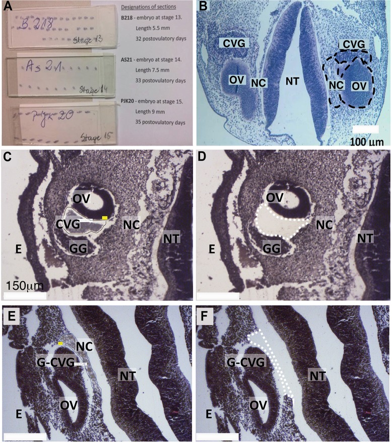
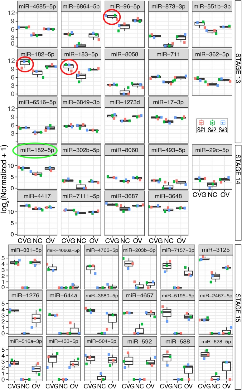
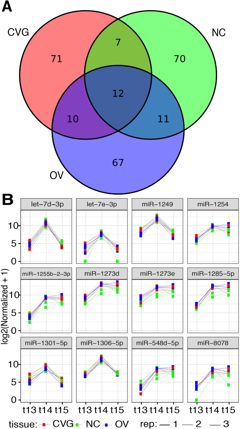
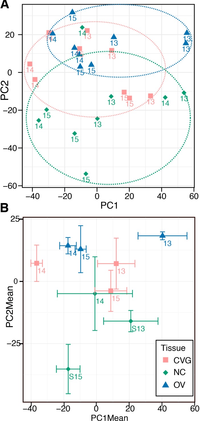
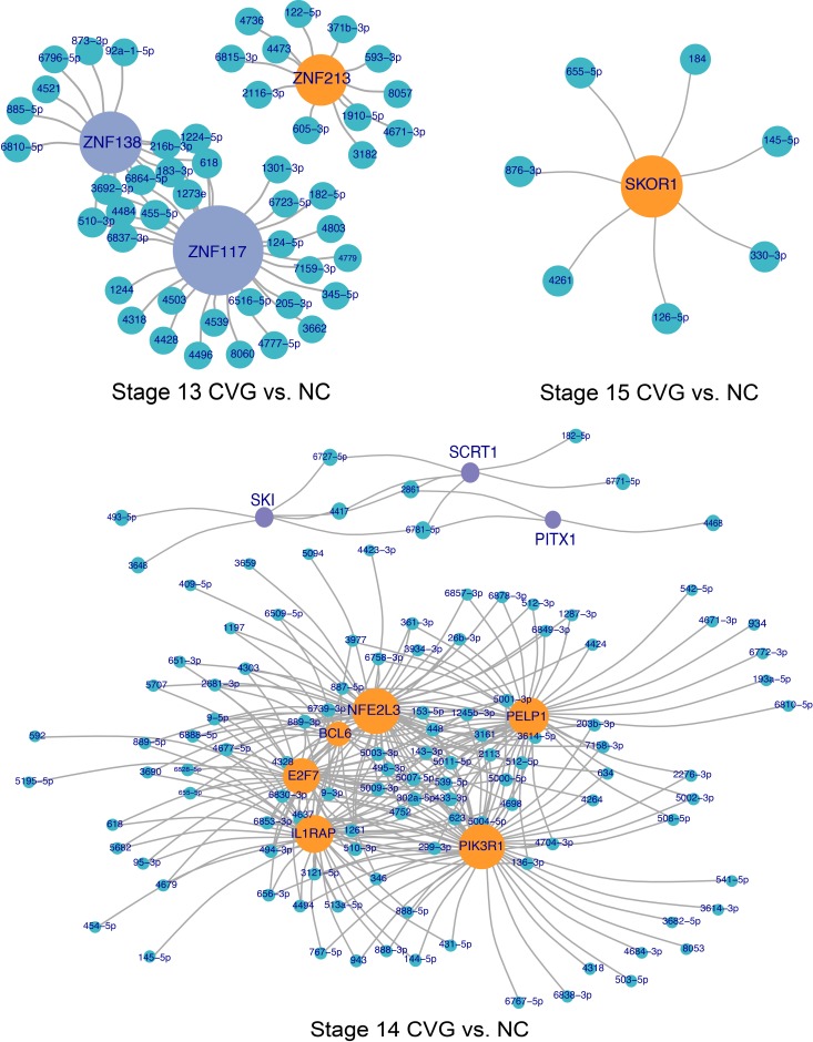
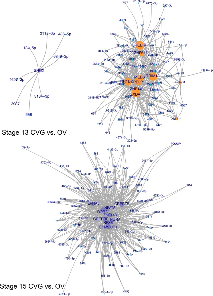
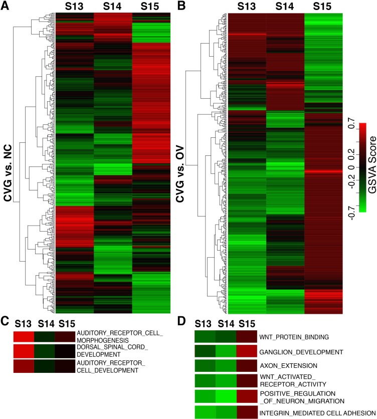
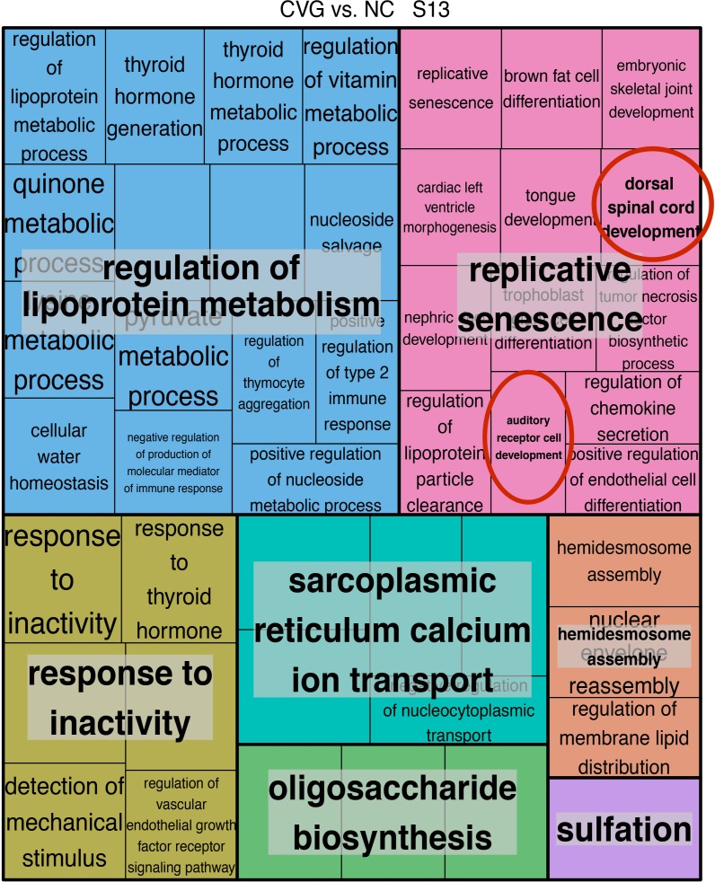
Due to the extreme inaccessibility of fetal human inner ear tissue, defining of the microRNAs (miRNAs) that regulate development of the inner ear has relied on animal tissue. In the present study, we performed the first miRNA sequencing of otic precursors in human specimens. Using HTG miRNA Whole Transcriptome assays, we examined miRNA expression in the cochleovestibular ganglion (CVG), neural crest (NC), and otic vesicle (OV) from paraffin embedded (FFPE) human specimens in the Carnegie developmental stages 13-15. We found that in human embryonic tissues, there are different patterns of miRNA expression in the CVG, NC and OV. In particular, members of the miR-183 family (miR-96, miR-182, and miR-183) are differentially expressed in the CVG compared to NC and OV at Carnegie developmental stage 13. We further identified transcription factors that are differentially targeted in the CVG compared to the other tissues from stages 13-15, and we performed gene set enrichment analyses to determine differentially regulated pathways that are relevant to CVG development in humans. These findings not only provide insight into the mechanisms governing the development of the human inner ear, but also identify potential signaling pathways for promoting regeneration of the spiral ganglion and other components of the inner ear.


Conflict of interest statement

Competing Interests: We have read the journal's policy and the authors of the manuscript have the following competing interests: MS, BL, and BJK are employees of HTG Molecular Diagnostics, Inc., which has developed HTG Edgeseq miRNA whole transcriptome assay used in this study. HTG Molecular Diagnostics, Inc. does not alter our adherence to PLOS ONE policies on sharing data and materials.

Figures

Fig 1
Fig 1. Laser-microdissection of FFPE-human inner ear slides.
(A) FFPE slides containing embryonic tissues. (B) Coronal section of a fetus with highlighted regions of interest (dashed black circles, CVG, NC, and OV). Further images are provided showing tissue before (C, E) and after (D, F) laser-captured microdissection of CVG (dashed white lines in D) and NC tissues (dashed white line in F). Other developmental regions also are highlighted. CVG: cochlear-vestibular ganglions, OV: otic vesicle, NC: neural crest; GG: geniculate ganglion, G-CVG: geniculate-cochleovestibular ganglions, E: epithelium; and NT: neural tube. Scale bar = 100 μm (B) and 150 μm (C-F).
Fig 2
Fig 2. Differential expression of miRNA across tissues.
Differentially expressed miRNA between three tissue types (CVG, NC, and OV) with p < 0.05 and false discovery adjusted p < 0.25, 0.50, or 0.10 (based on the smallest p-value out of the three possible tissue comparisons) are plotted for tissue from Carnegie stages 13, 14, and 15, respectively. MicroRNA from the miR-183 family is circled in red (stage 13) or green (stage 14).
Fig 3
Fig 3. Differentially expressed miRNA across time points.
(A) A Venn diagram is presented showing the top 100 miRNAs differentially expressed among the three time points (stages 13, 14, and 15) for each tissue type (CVG, NC, and OV). (B) Normalized counts are plotted for 12 commonly expressed miRNAs that are differentially expressed across time for all three tissues.
Fig 4
Fig 4. Principal component analysis.
(A) Principle components 1 and 2 are plotted for the 27 collected datasets. Loose clustering by tissue type is indicated with dashed colored circles. (B) As an alternative visualization, triplicate measures for each individual tissue/time point set are averaged and plotted with x- and y-standard error bars.
Fig 5
Fig 5. Transcription factor targeting network plots, CVG vs NC comparison.
Network plots showing differentially targeted transcription factors between CVG and NC tissues. Green nodes represent differentially expressed miRNAs, while orange and purple nodes represent significantly (false discovery adjusted p < 0.10) targeted transcription factors more targeted in either CVG (purple) or NC (orange) tissue. Transcription factor node size is proportional to the number of differentially expressed miRNAs targeting the gene.
Fig 6
Fig 6. Transcription factor targeting network plots, CVG vs OV comparison.
Network plots showing differentially targeted transcription factors between CVG and OV tissues. Green nodes represent differentially expressed miRNAs, while orange and purple nodes represent significantly (false discovery adjusted p < 0.10) targeted transcription factors more targeted in either CVG (purple) or OV (orange) tissue. Transcription factor node size is proportional to the number of differentially expressed miRNAs targeting the gene. Only results for stages 13 and 15 are shown, as there are too many significant factors to effectively visualize at stage 14.
Fig 7
Fig 7. Differentially activated gene ontological pathways by GSVA analysis.
Heatmap comparison of CVG with NC (A), and of CVG with OV (B). Each column represents the sample indicated at the top (three time points), each row represents an identified GO Term pathway (see the complete set in the S6 Dataset). The expression levels of GSVA scores are depicted according to the color scale (middle right). Red or green indicate expression levels above or below the median, respectively. The magnitude of deviation from the median is represented by color saturation. Heatmaps representing the GSVA score of GO term pathways relevant to CVG development that are significantly modulated in CVG. vs. NC comparison (C) and CVG vs. OV (D).
Fig 8
Fig 8. REVIGO treemap of relevance similarity analysis on enriched pathways comparing CVG vs. NC (stage 13).
REVIGO treemap summarizing Gene Ontology biological process categories over-represented in CVG cells compared to NC cells at stage 13. All terms are included with a FDR adjusted p-value cutoff at 0.05 from the enrichment analysis. The relevance similarity C-score (uniqueness) cut-off is chosen at 0.7. The size of each rectangle is proportional to the uniqueness for that category. Red circles indicate relevant CVG-development pathways.

Similar articles

Cited by

References

    1. Sayed D, Abdellatif M. MicroRNAs in development and disease. Physiol Rev. 2011;91: 817–887. doi: 10.1152/physrev.00006.2010 MICRORNAS - DOI - PubMed
    1. Shenoy A, Blelloch RH. Regulation of microRNA function in somatic stem cell proliferation and differentiation. Nat Rev Mol Cell Biol. Nature Publishing Group; 2014;15: 565–76. doi: 10.1038/nrm3854 - DOI - PMC - PubMed
    1. Weston MD, Pierce ML, Rocha-Sanchez S, Beisel KW, Soukup G a. MicroRNA gene expression in the mouse inner ear. Brain Res. 2006;1111: 95–104. doi: 10.1016/j.brainres.2006.07.006 - DOI - PubMed
    1. Soukup GA, Fritzsch B, Pierce ML, Weston MD, Jahan I, McManus MT, et al. Residual microRNA expression dictates the extent of inner ear development in conditional Dicer knockout mice. Dev Biol. Elsevier Inc.; 2009;328: 328–341. doi: 10.1016/j.ydbio.2009.01.037 - DOI - PMC - PubMed
    1. Friedman LM, Dror A a, Avraham KB. Mouse models to study inner ear development and hereditary hearing loss. Int J Dev Biol. 2007;51: 609–31. doi: 10.1387/ijdb.072365lf - DOI - PubMed

Publication types

MeSH terms