. 2018 Jan 26;13(1):e0191916.
doi: 10.1371/journal.pone.0191916. eCollection 2018.

Modulation of HIV replication in monocyte derived macrophages (MDM) by steroid hormones

Affiliations

Modulation of HIV replication in monocyte derived macrophages (MDM) by steroid hormones

Krishnakumar Devadas et al. PLoS One. .

Abstract

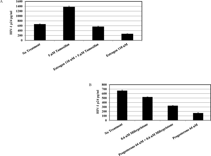
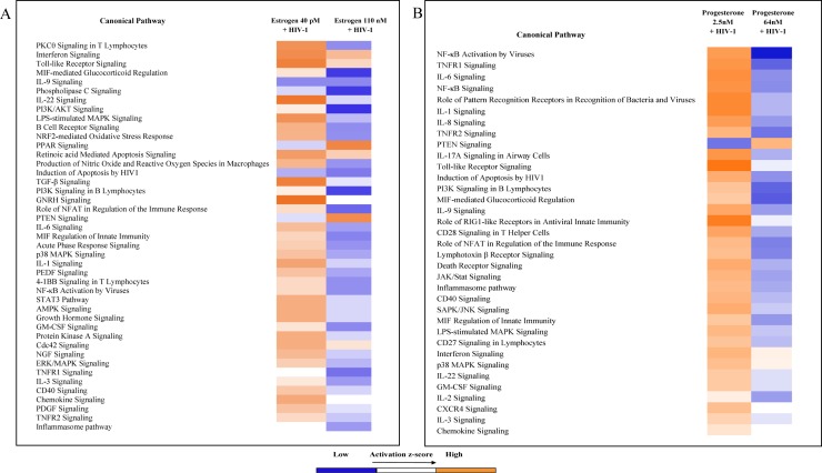
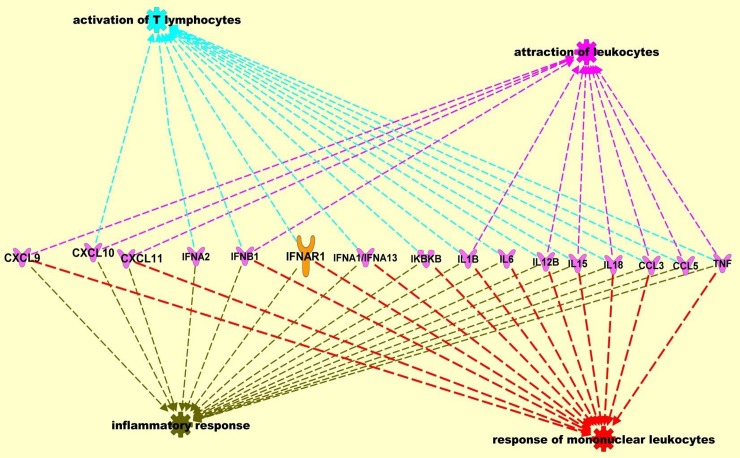
Significant sex specific differences in the progression of HIV/AIDS have been reported. Several studies have implicated steroid hormones in regulating host factor expression and modulating HIV transmission and replication. However, the exact mechanism exerted by steroid hormones estrogen and progesterone in the regulation of HIV-1 replication is still unclear. Results from the current study indicated a dose dependent down regulation of HIV-1 replication in monocyte derived macrophages pre-treated with high concentrations of estrogen or progesterone. To elucidate the molecular mechanisms associated with the down regulation of HIV-1 replication by estrogen and progesterone we used PCR arrays to analyze the expression profile of host genes involved in antiviral responses. Several chemokines, cytokines, transcription factors, interferon stimulated genes and genes involved in type-1 interferon signaling were down regulated in cells infected with HIV-1 pre-treated with high concentrations of estrogen or progesterone compared to untreated HIV-1 infected cells or HIV-1 infected cells treated with low concentrations of estrogen or progesterone. The down regulation of CXCL9, CXCL10 and CXCL11 chemokines and IL-1β, IL-6 cytokines in response to high concentrations of estrogen and progesterone pre-treatment in HIV-1 infected cells was confirmed at the protein level by quantitating chemokine and cytokine concentrations in the culture supernatant. These results demonstrate that a potent anti-inflammatory response is mediated by pre-treatment with high concentrations of estrogen and progesterone. Thus, our study suggests a strong correlation between the down-modulation of anti-viral and pro-inflammatory responses mediated by estrogen and progesterone pre-treatment and the down regulation of HIV-1 replication. These findings may be relevant to clinical observations of sex specific differences in patient populations and point to the need for further investigation.


Conflict of interest statement

Competing Interests: The authors have declared that no competing interests exist.

Figures

Fig 1
Fig 1. Modulation of HIV-1 replication by estrogen and progesterone.
MDMs (1x10 6 cells/well) were pre-treated with appropriate concentrations of estrogen or progesterone and infected with HIV-1. After 2 hours the virus was removed, and fresh culture media added with appropriate hormones and cultured. Hormone concentrations used: Estrogen: 1.75 μM, 110 nM, 140 pM, 40 pM; Progesterone: 64 nM, 32 nM, 2.5 nM and 1 pM. Culture supernatants were collected 9 days and / or 12 days post infection and HIV-1 replication quantitated by p24 ELISA. Culture supernatants were analyzed in triplicate. Results expressed as mean ± SEM are representative of three independent experiments. Asterisk (*) over the bars indicates significant difference with control, *** P <0.0001, ** P <0.001 and * P <0.05. P-values were generated by Student’s t-test and comparisons were done with control. A. MDMs infected with HIV-1 BaL at 5 ng/ml and pre-treated with estrogen. B. MDMs infected with HIV-1 BaL at 5 ng/ml and pre-treated with progesterone. C. MDMs infected with HIV-1 Clade B NSI at 5 ng/ml and pretreated with estrogen. D. MDMs infected with HIV-1 Clade B NSI at 5 ng/ml and pre-treated with progesterone. E. MDMs infected with HIV-1 Clade C NSI at 5 ng/ml and pretreated with estrogen. F. MDMs infected with HIV-1 Clade C NSI at 5 ng/ml and pre-treated with progesterone.
Fig 2
Fig 2. HIV-1 BaL replication in MDMs treated with mifepristone and tamoxifen.
MDMs (1 x10 6 cells/well) were pre-treated with concentrations of 5 μM tamoxifen or 0.6 nM mifepristone for 2 hours and appropriate concentrations of estrogen or progesterone were added and cultured for 5 days. The cells were infected with HIV-1 BaL at 5 ng/ml and cultured in media containing 110 nM estrogen and 64 nM progesterone. Culture supernatants were harvested 9 days post infection and HIV-1 replication quantitated by p24 ELISA. Culture supernatants were analyzed in triplicate. Results expressed as mean ± SEM are representative of three independent experiments.A. Tamoxifen and estrogen pre-treated MDMs B. Mifepristone and progesterone pre-treated MDMs.
Fig 3
Fig 3. Identification of differentially expressed genes in estrogen or progesterone treated MDMs infected with HIV-1 using RT2 Profiler PCR Arrays.
Assays were performed with experimental RNA samples isolated from MDMs obtained from 3 independent donors. A. Differentially expressed genes in estrogen treated MDMs infected with HIV-1 BaL.B. Differentially expressed genes in progesterone treated cells.
Fig 4
Fig 4. BioPlex analysis of secreted cytokines and chemokines in culture supernatants.
A. BioPlex analysis of secreted cytokines and chemokines in culture supernatants from monocytes derived macrophage cells infected with HIV-1 (BaL) pre-treated with 40 pM or 110 nM estrogen and MDMs not infected with HIV-1, not pre-treated with estrogen as control. Presented in this panel are Tumor necrosis factor-α (TNF-α), Interferon-α2 (IFN-α2), Chemokine (C-X-C motif) ligand 9 (CXCL9), Chemokine (C-X-C motif) ligand 10 (CXCL10), Chemokine (C-X-C motif) ligand 11 (CXCL11), Interleukin 1 β (IL1β), Interleukin-6 (IL-6), Interleukin-18 (IL-18). Culture supernatants were analyzed in duplicate. Results expressed as mean ± SEM are representative of three independent experiments. B. BioPlex analysis of secreted cytokines and chemokines in culture supernatants from monocytes derived macrophage cells infected with HIV-1 (BaL) pre-treated with 2.5 nM or 64 nM progesterone and MDMs not infected with HIV-1, not pre-treated with progesterone as control. Presented in this panel are Tumor necrosis factor-α (TNF-α), Interferon-α2 (IFN-α2), Chemokine (C-X-C motif) ligand 9 (CXCL9), Chemokine (C-X-C motif) ligand 10 (CXCL10), Chemokine (C-X-C motif) ligand 11 (CXCL11), Interleukin 1 β (IL1β), Interleukin-6 (IL-6), Interleukin-18 (IL-18). Culture supernatants were analyzed in duplicate. Results expressed as mean ± SEM are representative of three independent experiments.
Fig 5
Fig 5. Ingenuity Pathway Analysis (IPA) of the differentially expressed host genes identified by the PCR array.
Data sets were analyzed through the use of QIAGEN's Ingenuity® Pathway Analysis (IPA®, QIAGEN Redwood City, CA, USA www.qiagen.com/ingenuity). Identification of the canonical pathways from Ingenuity Pathways Knowledge Base (IPKB) most significantly associated with the genes differentially expressed between MDMs infected with HIV-1 pre-treated with estrogen (A) or progesterone (B) and MDMs infected with HIV-1 not treated with estrogen or progesterone samples. The differentially expressed genes identified by the PCR array were used for the analysis. The significance of the association was measured on the basis of the ratio of the number of genes from the data set that map to the pathway divided by the total number of genes that map to the canonical pathway (as displayed); and a p-value determining the probability that the association between the genes in the data set and the canonical pathway is explained by chance alone (Fischer’s exact test).
Fig 6
Fig 6. Ingenuity Pathway Analysis (IPA) of the differentially expressed host genes identified by the PCR array.
Data sets were analyzed through the use of QIAGEN's Ingenuity® Pathway Analysis (IPA®, QIAGEN Redwood City, CA, USA www.qiagen.com/ingenuity). Network analysis included direct and indirect relationships with disease functions and selected genes differentially expressed between MDMs infected with HIV-1 pre-treated with estrogen or progesterone and MDMs infected with HIV-1 not treated with estrogen or progesterone samples.

Similar articles

Cited by

References

    1. Long EM, Martin HL Jr., Kreiss JK, Rainwater SM, Lavreys L, Jackson DJ, et al. Gender differences in HIV-1 diversity at time of infection. Nat Med. 2000;6(1):71–5. Epub 1999/12/29. doi: 10.1038/71563 . - DOI - PubMed
    1. Anastos K, Gange SJ, Lau B, Weiser B, Detels R, Giorgi JV, et al. Association of race and gender with HIV-1 RNA levels and immunologic progression. J Acquir Immune Defic Syndr. 2000;24(3):218–26. Epub 2000/09/02. . - PubMed
    1. Gandhi M, Bacchetti P, Miotti P, Quinn TC, Veronese F, Greenblatt RM. Does patient sex affect human immunodeficiency virus levels? Clin Infect Dis. 2002;35(3):313–22. Epub 2002/07/13. doi: CID011586 [pii] doi: 10.1086/341249 . - DOI - PubMed
    1. Klein SL. The effects of hormones on sex differences in infection: from genes to behavior. Neurosci Biobehav Rev. 2000;24(6):627–38. . - PubMed
    1. Fish EN. The X-files in immunity: sex-based differences predispose immune responses. Nat Rev Immunol. 2008;8(9):737–44. doi: 10.1038/nri2394 . - DOI - PMC - PubMed

Publication types