. 2019 Feb 1;29(2):701-715.
doi: 10.1093/cercor/bhx353.

Fear Extinction Recall Modulates Human Frontomedial Theta and Amygdala Activity

Affiliations

Fear Extinction Recall Modulates Human Frontomedial Theta and Amygdala Activity

Matthias F J Sperl et al. Cereb Cortex. .

Abstract

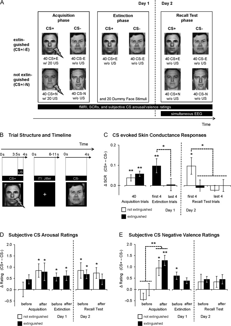
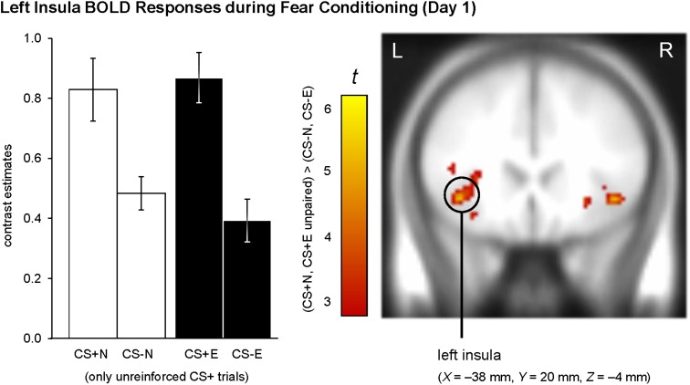
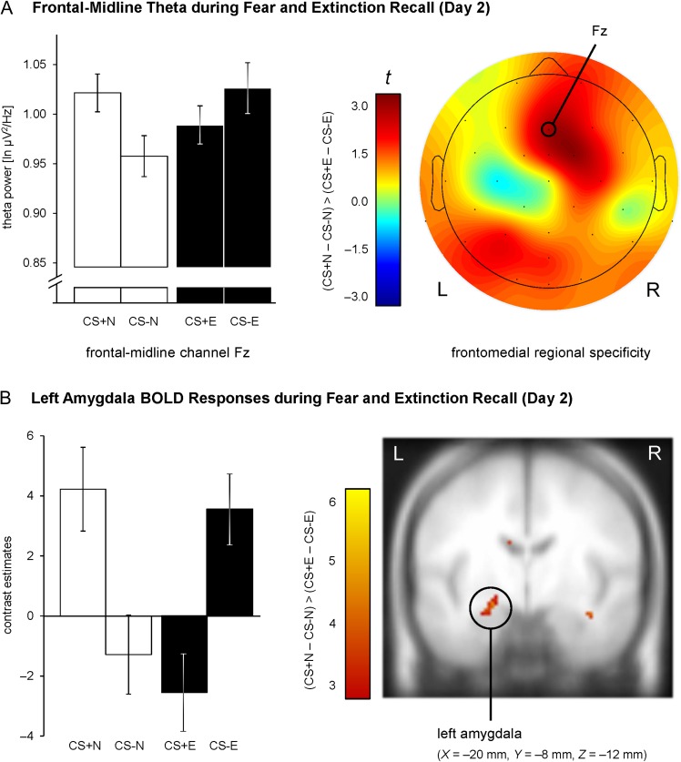
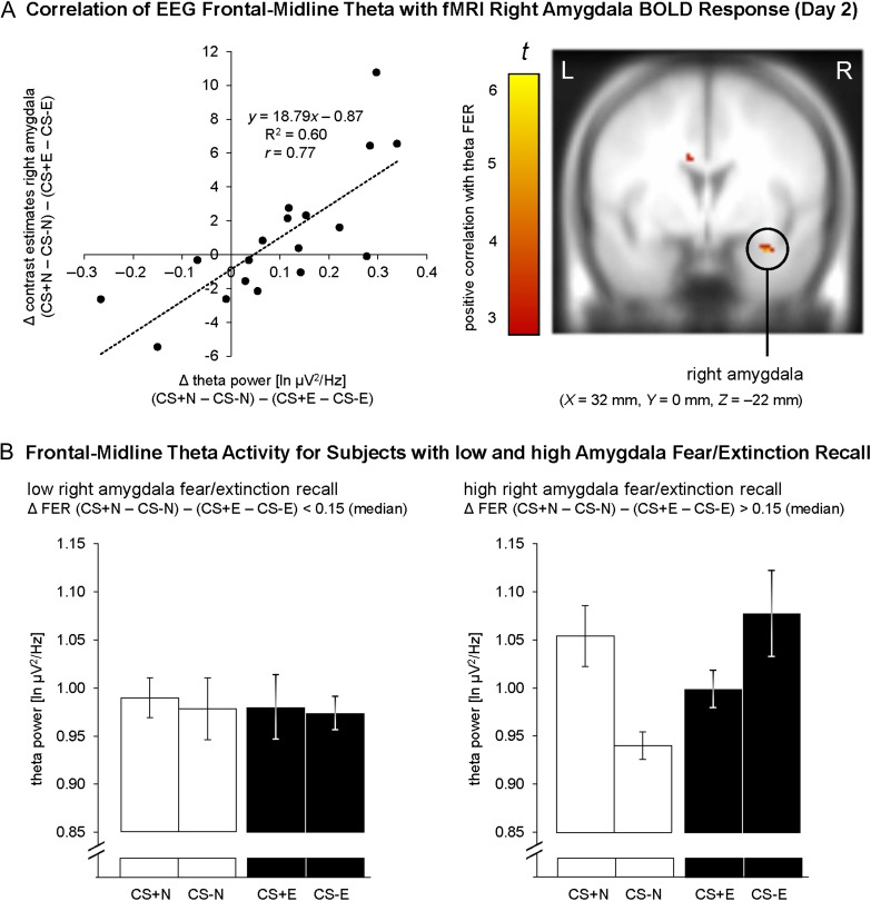
Human functional magnetic resonance imaging (fMRI) and electroencephalography (EEG) studies, as well as animal studies, indicate that the amygdala and frontomedial brain regions are critically involved in conditioned fear and that frontomedial oscillations in the theta range (4-8 Hz) may support communication between these brain regions. However, few studies have used a multimodal approach to probe interactions among these key regions in humans. Here, our goal was to bridge the gap between prior human fMRI, EEG, and animal findings. Using simultaneous EEG-fMRI recordings 24 h after fear conditioning and extinction, conditioned stimuli presented (CS+E, CS-E) and not presented during extinction (CS+N, CS-N) were compared to identify effects specific to extinction versus fear recall. Differential (CS+ vs. CS-) electrodermal, frontomedial theta (EEG) and amygdala responses (fMRI) were reduced for extinguished versus nonextinguished stimuli. Importantly, effects on theta power covaried with effects on amygdala activation. Fear and extinction recall as indicated by theta explained 60% of the variance for the analogous effect in the right amygdala. Our findings show for the first time the interplay of amygdala and frontomedial theta activity during fear and extinction recall in humans and provide insight into neural circuits consistently linked with top-down amygdala modulation in rodents.


Figures

Figure 1.
Figure 1.
Schematic depiction of the experimental paradigm used in the present study. (A) Number and stimuli types presented during the 3 experimental phases. The central hypotheses of the current study focused on the Day 2 recall test, during which EEG and fMRI were recorded simultaneously. CS+E/CS−E, CSs presented during extinction phase; CS+N/CS−N, CSs not presented during extinction phase. CS+ were reinforced with an aversive US (“w/”, contingency of 50%) during acquisition phase, while CS− were never paired with a US (“w/o”). (B) Trial structure and timeline for a single CS trial. All CSs were shown for 4 s. During the acquisition phase, a 500-ms electric shock US coterminated with 50% of all CS+ trials, starting 3.5 s after CS onset. (C) Normalized CS evoked differential (CS+ – CS−) SCRs, (D) subjective CS arousal ratings, and (E) subjective CS negative valence ratings (M ± within-subject standard error of the mean (SEM), O'Brien and Cousineau 2014) for extinguished and nonextinguished stimuli during all experimental stages. *P ≤ 0.05, **P ≤ 0.01 (one-sided, CS+ > CS−).
Figure 2.
Figure 2.
fMRI correlates of fear conditioning on Day 1. Insula activation was significantly enhanced for CS+ (CS+E, CS+N) compared with CS− (CS−E, CS−N). To confirm that neural responses did not differ between to-be extinguished and to-be nonextinguished stimuli, we constructed an additional first-level GLM which contained separate regressors for to-be extinguished and to-be nonextinguished CS+/CS−. Contrast estimates were extracted and subjected to a Contingency × Later Extinction Status ANOVA, which did not show a significant interaction, F(1,17) = 0.65, P = 0.431, but confirmed a significant main effect for Contingency, F(1,17) = 24.45, P < 0.001. For illustration purposes, the intensity threshold was set to P ≤ 0.005 (uncorrected) with a minimal cluster threshold of k ≥ 5 contiguous significant voxels. Activations (t-values) were superimposed on the MNI305 T1 template. All coordinates (X, Y, Z) are given in MNI space. L = left, R = right brain hemisphere. Bar graphs show the mean contrast estimates (±within-subject SEM, O'Brien and Cousineau 2014) for a cluster of voxels with P ≤ 0.005 (uncorrected) surrounding the peak voxel within the insula ROI.
Figure 3.
Figure 3.
EEG and fMRI correlates of fear and extinction recall on Day 2. (A) Differential (CS+ – CS−) ln-transformed theta power at frontal-midline channel Fz was significantly reduced for extinguished versus nonextinguished stimuli (left). This effect was specific for frontomedial electrode channels (right). Bar graphs show the mean theta power (± within-subject SEM, O'Brien and Cousineau 2014). (B) Reduced differential amygdala responses (CS+ – CS−) for extinguished compared with nonextinguished stimuli. Habituation of amygdala activity was modeled by an exponentially decaying function, based on habituation of SCRs. For illustration purposes, the intensity threshold was set to P ≤ 0.005 (uncorrected) with a minimal cluster threshold of k ≥ 5 contiguous significant voxels. Activations (t-values) were superimposed on the MNI305 T1 template. All coordinates (X, Y, Z) are given in MNI space. L = left, R = right brain hemisphere. Bar graphs show the mean contrast estimates (± within-subject SEM, O'Brien and Cousineau 2014) for a cluster of voxels with P ≤ 0.005 (uncorrected) surrounding the peak voxel within the amygdala ROI.
Figure 4.
Figure 4.
Integration of EEG frontomedial (Fz) theta power and fMRI right amygdala activation of fear and extinction recall on Day 2. (A) Positive correlation of theta modulations to conditioned and extinguished fear with BOLD responses in the right amygdala. Consistent with our assumed involvement of theta oscillations in AMC-amygdala connectivity (Gilmartin et al. 2014), this correlation indicates that subjects with relatively strong amygdala activation to nonextinguished (vs. extinguished) fear stimuli are characterized by relatively strong differential frontomedial theta power. For illustration purposes, the intensity threshold was set to P ≤ 0.005 (uncorrected) with a minimal cluster threshold of k ≥ 5 contiguous significant voxels. Activations (t-values) were superimposed on the MNI305 T1 template. All coordinates (X, Y, Z) are given in MNI space. L = left, R = right brain hemisphere. (B) To illustrate the positive correlation, right amygdala BOLD responses for the FER contrast (CS+N – CS−N) – (CS+E – CS−E) were compared based on median split, and theta power was assessed separately for subjects with low and high amygdala fear/extinction recall, that is, low/high FER BOLD scores (bar graphs show M ± within-subject SEM, O'Brien and Cousineau 2014). Higher differential theta power for nonextinguished versus extinguished CSs only emerged for subjects with high (P < 0.001), but not with low (P = 0.929) fear/extinction recall in the right amygdala.

References

    1. Allen PJ, Josephs O, Turner R. 2000. A method for removing imaging artifact from continuous EEG recorded during functional MRI. NeuroImage. 12:230–239. - PubMed
    1. Allen PJ, Polizzi G, Krakow K, Fish DR, Lemieux L. 1998. Identification of EEG events in the MR scanner: the problem of pulse artifact and a method for its subtraction. NeuroImage. 8:229–239. - PubMed
    1. Amano T, Duvarci S, Popa D, Pare D. 2011. The fear circuit revisited: contributions of the basal amygdala nuclei to conditioned fear. J Neurosci. 31:15481–15489. - PMC - PubMed
    1. Armony JL, Dolan RJ. 2001. Modulation of auditory neural responses by a visual context in human fear conditioning. Neuroreport. 12:3407–3411. - PubMed
    1. Bastiaansen M, Mazaheri A, Jesen O. 2012. Beyond ERPs: oscillatory neuronal dynamics In: Luck SJ, Kappenman ES, editors. The Oxford handbook of event-related potential components. New York, NY: Oxford University Press. p. 31–49.

Publication types