Comparative Study
. 2018 May 1;35(5):1047-1062.
doi: 10.1093/molbev/msx336.

Whole-Body Single-Cell Sequencing Reveals Transcriptional Domains in the Annelid Larval Body

Affiliations
Comparative Study

Whole-Body Single-Cell Sequencing Reveals Transcriptional Domains in the Annelid Larval Body

Kaia Achim et al. Mol Biol Evol. .

Abstract

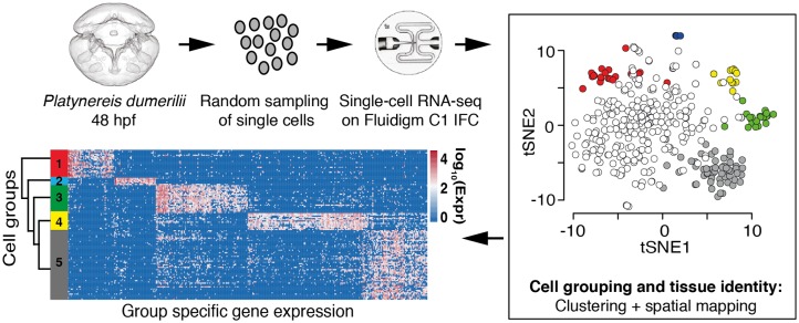
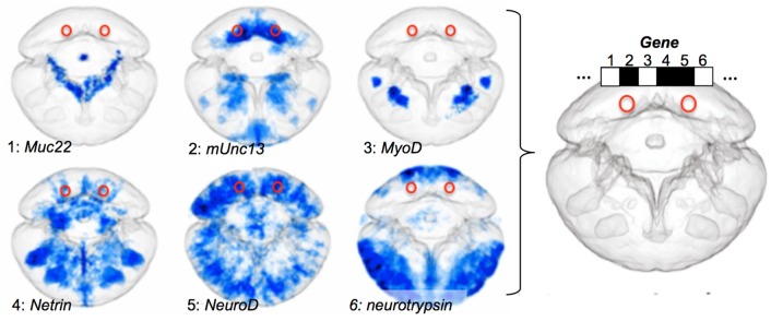
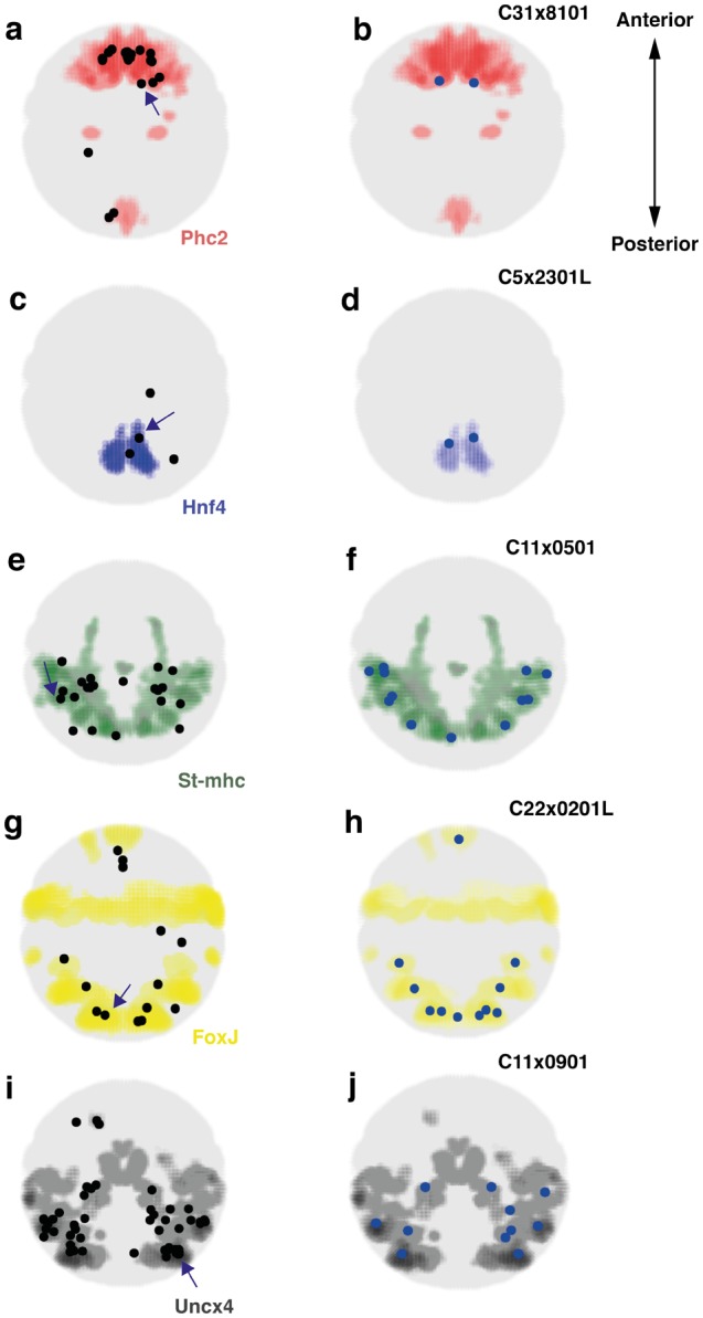
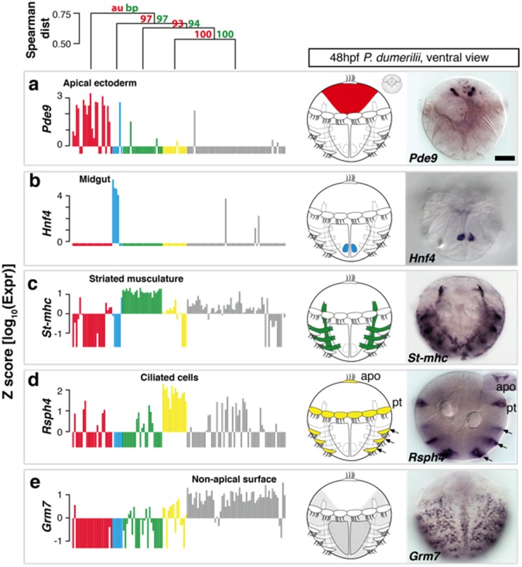
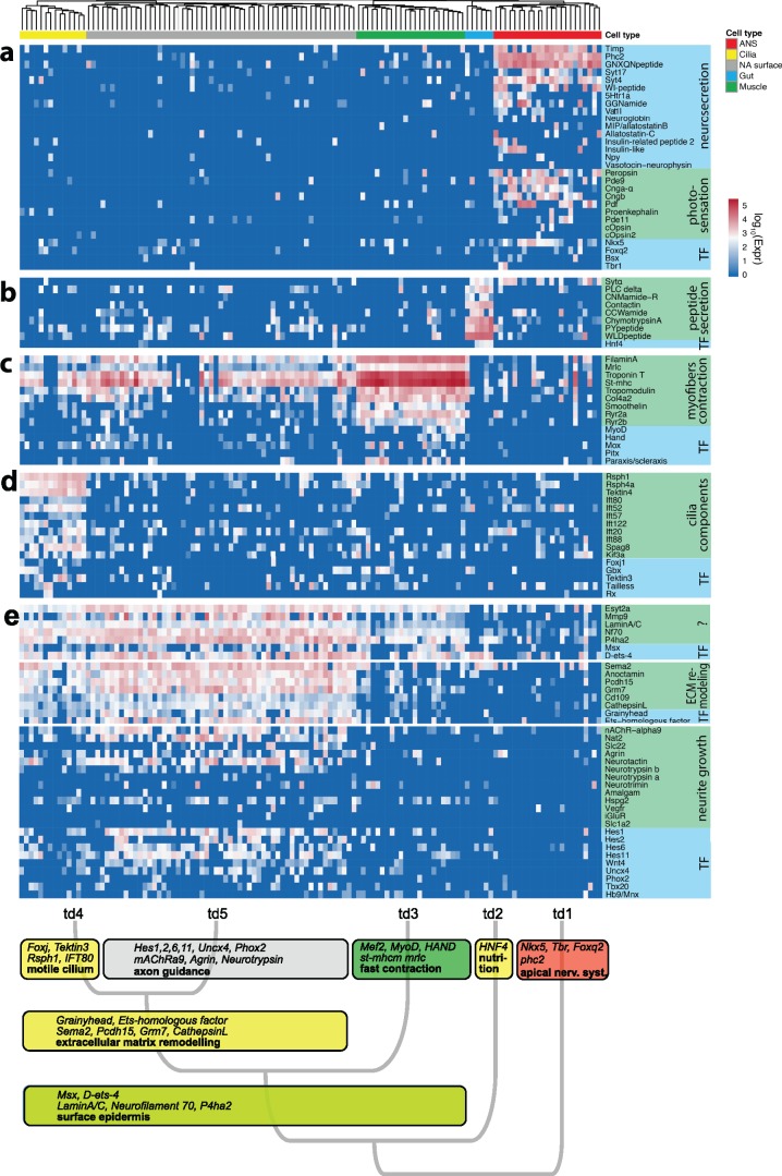
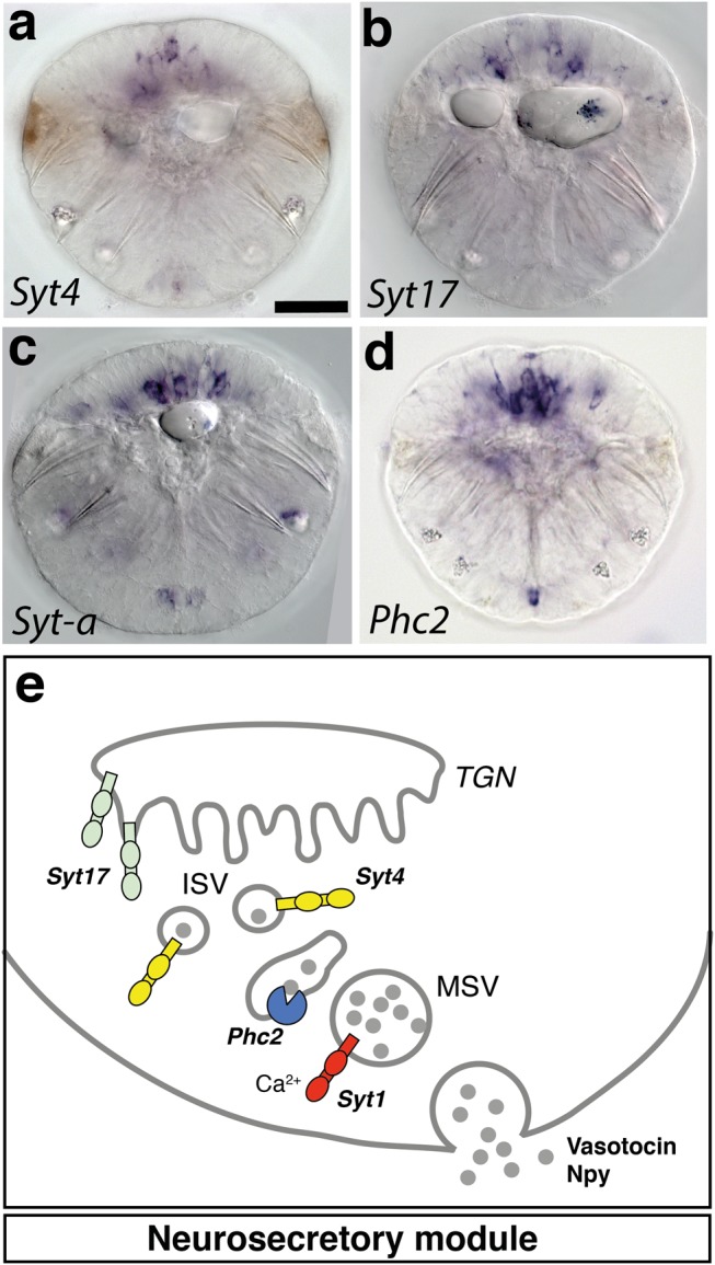
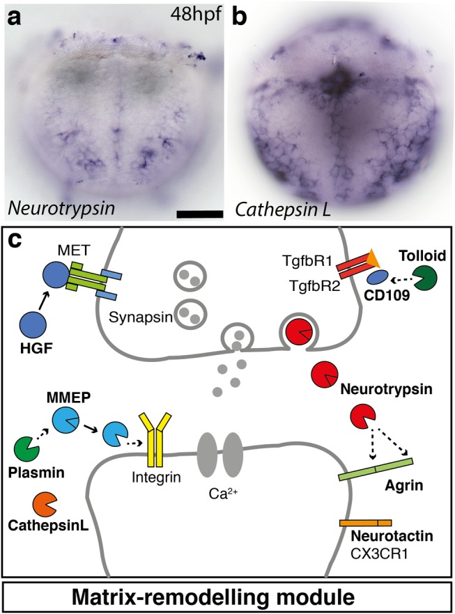
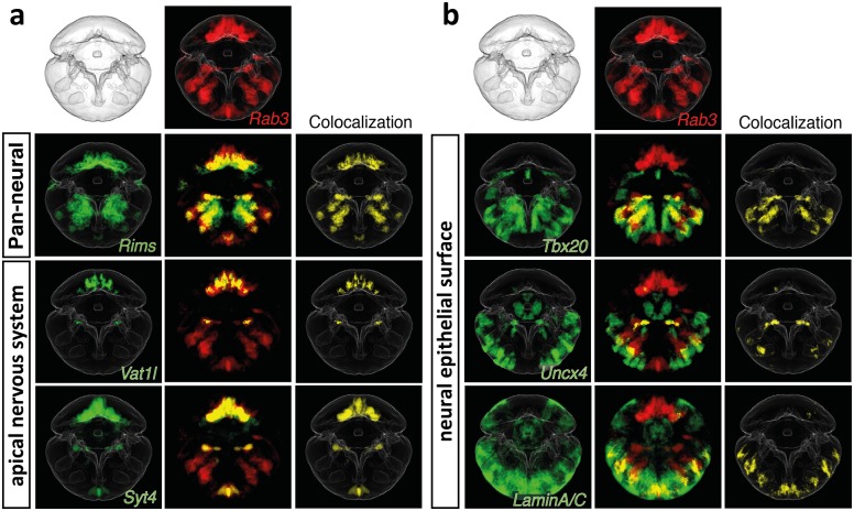
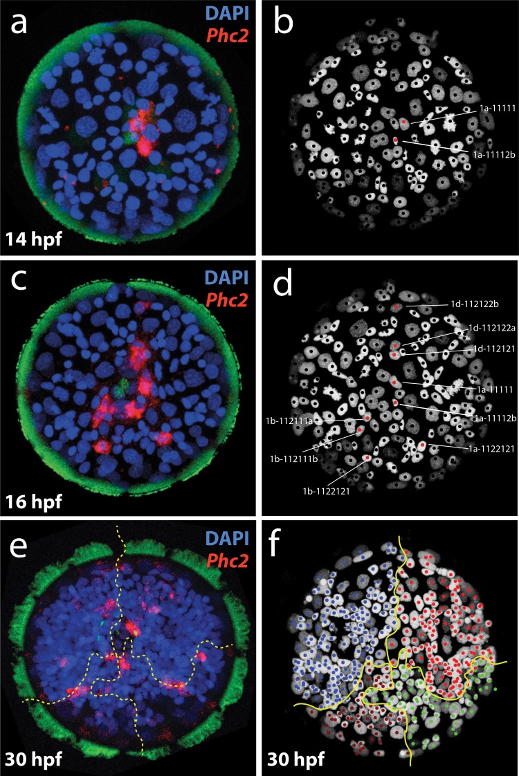
Animal bodies comprise diverse arrays of cells. To characterize cellular identities across an entire body, we have compared the transcriptomes of single cells randomly picked from dissociated whole larvae of the marine annelid Platynereis dumerilii. We identify five transcriptionally distinct groups of differentiated cells, each expressing a unique set of transcription factors and effector genes that implement cellular phenotypes. Spatial mapping of cells into a cellular expression atlas, and wholemount in situ hybridization of group-specific genes reveals spatially coherent transcriptional domains in the larval body, comprising, for example, apical sensory-neurosecretory cells versus neural/epidermal surface cells. These domains represent new, basic subdivisions of the annelid body based entirely on differential gene expression, and are composed of multiple, transcriptionally similar cell types. They do not represent clonal domains, as revealed by developmental lineage analysis. We propose that the transcriptional domains that subdivide the annelid larval body represent families of related cell types that have arisen by evolutionary diversification. Their possible evolutionary conservation makes them a promising tool for evo-devo research.


Figures

<sc>Fig</sc>. 1.
Fig. 1.
Single-cell transcriptomics of Platynereis 48 hpf larvae. Cells of the 48 hpf larvae were dissociated and randomly selected for single-cell RNA-sequencing using the Fluidigm C1 Single-cell AutoPrep system. Combining sparse clustering with spatial positioning of single cells allows the identification of robust cell groups within the data. The clustering approach enables identification of genes that characterize each cell type. Finally, we used hierarchical clustering to investigate the similarity between the identified cell clusters.
<sc>Fig</sc>. 2.
Fig. 2.
The ProSPr cellular expression atlas for 48 hpf Platynereis larvae. ProSPr generates a standardized expression pattern for each gene (1–6 are examples) that are registered onto the same averaged larva. As a result, for any bilateral pair of cells in the body (red rings) the expression profile can be determined in a binary fashion (white: not expressed; black: expressed).
<sc>Fig</sc>. 3.
Fig. 3.
Spatial mapping of single cells to spatially coherent regions characterized by specific marker gene expression. Expression of specific marker gene expression visualized as colored regions within the larva in ventral views. Left panels: Mapping results for all cells belonging to the respective group. Centroids of the voxel clusters to which cells mapped with highest confidence are shown as black dots. Right panels: For each cell group, we show an example of the mapping result for one individual cell (indicated by an arrow on the left panel). (a) Cells of the apical ectoderm group (n=23 cells) map to the Phc2 expressing region in the embryo. (b) Mapping result for the cell C31x8101. (c) Midgut cells (n=4 cells) map to the Hnf4 expressing region. (d) Mapping result for the cell C5x2301L. (e) Striated musculature cells (n=23 cells) map to the St-mhc expressing region. (f) Mapping result for the cell C11x0501. (g) Cells of the ciliary bands (n=14 cells) map to the Foxj expressing region. (h) Mapping result for the cell C22x0201L. (i) Cells of the nonapical ectodermal group (n=55 cells) map to the Uncx4 expression domain. (j) Mapping result for the cell C11x0901.
<sc>Fig</sc>. 4.
Fig. 4.
Identification and validation of tissue-specific genes. On top, Distance tree showing the hierarchical relationships between the differentiated cell groups. (a–e) For each identified cell group, the expression of a group-specific marker gene is shown in a bar plot, the respective tissue is shown schematically in the ventral view of Platynereis larva, and visualized by WMISH with respective probes: (a) Pde9 expression in the apical ectoderm (red); (b) Hnf4 expression in the midgut (cyan); (c) St-mhc expression in striated muscle (green); (d) Rsph4 expression in ciliated cells (yellow); and (e) Grm7 expression characterizes the nonapical surface cells (gray). Note that Pde9, Hnf4, Rsph4 and Grm7 are novel markers for the respective cell groups. Each ISH pattern was replicated in at least six animals. Scale bar, 50 μm. Apo, apical organ; pt, prototroch, Z factor=xij-μiσi, xij, expression of gene i in cell j, μi, mean expression of gene i, σi, SD of gene i.
<sc>Fig</sc>. 5.
Fig. 5.
Spatial extent of the group 1 versus group 5 transcriptional domains as determined by specific marker gene comparison. Gene expression averages at 48 hpf as determined by the ProSPr cellular expression atlas. Group 1 gene Phc2 in red; group 2 genes LaminAC and Pcdh15 in green. (a and b) Apical views; (c and d) ventral views. The inset in (a) and (b) is a scanning electron microscopy apical view in the same orientation, illustrating that the group 1 domains is situated around the apical organ.
<sc>Fig</sc>. 6.
Fig. 6.
Tissue specific marker genes reflect cellular functions. For each of the differentiated tissues, we show a heatmap of tissue specific gene expression: (a) expression profile of group 1 apical nervous system-specific genes; (b) expression profile of group 2 midgut genes; (c) expression profile of group 3 myocyte-specific genes; (d) expression profile of group 4 multiciliated cell-specific genes; and (e) expression profile of group 5 neural/epithelial surface-specific genes. Functionally related groups of genes are highlighted. TF: transcription factor, td: transcriptional domain.
<sc>Fig</sc>. 7.
Fig. 7.
Neurosecretory functional module is specific to group 1 ANS cells. (a–d) WMISH analysis of Syt4, Syt17, Sytα, and Phc2 expression in Platynereis larvae at 48 hpf. Ventral view. Scale bar, 50 μm. (e) Schematic of the neurosecretion cellular module. Syt17 contains an N-terminal cysteine cluster mediating membrane association with the trans-Golgi network (TGN) (Fukuda 2003). Syt4 is involved in the maturation of secretory vesicles (Zhang et al. 2011). Sytα functions in vesicle trafficking of specific subclasses of neuropeptides and/or neuromodulators (Adolfsen et al. 2004). TGN, trans-Golgi network; ISV, immature secretory vesicle, MSV, mature secretory vesicle.
<sc>Fig</sc>. 8.
Fig. 8.
Extracellular matrix remodeling module is specific to group 5 neural/epithelial surface cells. (a and b) WMISH analysis of Neurotrypsin and Cathepsin L expression in Platynereis larvae at 48 hpf. Ventral view focused at the surface. Scale bar, 50 μm c, Schematic of the synapse formation cellular module. Neurotrypsin cleaves agrin locally at the synaptic cleft, triggering the formation of new synapses (Sonderegger and Matsumoto-Miyai 2014). HGF and its receptor MET enhance clustering of synaptic proteins at excitatory synapses (Tyndall and Walikonis 2006). Plasmin cleaves selected synaptic target proteins such as NMDA receptor and matrix metalloproteinases (Sonderegger and Matsumoto-Miyai 2014). Several members of the family of matrix-metalloproteinases such as MMP-9 have been implicated in synapse formation and remodeling (Michaluk et al. 2011). Tolloid-like (Tll) and its substrate CD109 have been implicated in the control of TGFβ signaling and extracellular matrix synthesis (Vadon-Le Goff et al. 2015). Cathepsin L is likewise implicated in extracellular matrix remodeling and neuronal survival (Felbor et al. 2002). The genes specifically expressed in nonapical ectodermal cells are marked in bold.
<sc>Fig</sc>. 9.
Fig. 9.
Coexpression of groups 1 and 5 marker genes with the pan-neuronal marker Rab3 indicates the presence of neural cell types. Averaged gene expression patterns for 48 hpf ProSPr used for in silico analysis of coexpression. (a) Group 1. (b) Group 5.
<sc>Fig</sc>. 10.
Fig. 10.
The developmental lineage of early differentiating Phc2+ cells. (a–d) Multiclonal origin of the first 10 differentiating Phc2+ cells. For each labeled cell, the upper line indicates the name of the cell and the lower line its lineage progeny. (a and b) Represent 14 hpf and (c and d) represent 16 hpf. (a and c) Confocal images of Phc2+ cells and (b and d) the corresponding cells in the tracked lineage (Vopalensky et al. 2018) indicated by red dots. Note that the 10 phc2+ cells labeled in (d) are of different clonal origin as revealed by their deviating lineage progeny. (e and f) The cells present at 30 hpf also mostly originate from different clonal domains, as revelaed by spatial alignment to the clonal map at 30 hpf. Brown lines separate cells descending from the first four distinct cells A, B, C, D.

References

    1. Achim K, Pettit J-B, Saraiva LR, Gavriouchkina D, Larsson T, Arendt D, Marioni JC.. 2015. High-throughput spatial mapping of single-cell RNA-seq data to tissue of origin. Nat Biotechnol. 335:503–509. - PubMed
    1. Ackermann C, Dorresteijn A, Fischer A.. 2005. Clonal domains in postlarval Platynereis dumerilii (Annelida: Polychaeta). J Morphol. 2663:258–280. - PubMed
    1. Adolfsen B, Saraswati S, Yoshihara M, Littleton JT.. 2004. Synaptotagmins are trafficked to distinct subcellular domains including the postsynaptic compartment. J Cell Biol. 1662:249–260. - PMC - PubMed
    1. Anders S, Pyl PT, Huber W.. 2015. HTSeq–a Python framework to work with high-throughput sequencing data. Bioinformatics 312:166–169.http://dx.doi.org/10.1093/bioinformatics/btu638 - DOI - PMC - PubMed
    1. Andrews S. 2010. FastQC: a quality control tool for high throughput sequence data. Available online at:http://www.bioinformatics.babraham.ac.uk/projects/fastqc/

Publication types

LinkOut - more resources