Clinical Trial
. 2018 Apr 1;27(7):1228-1240.
doi: 10.1093/hmg/ddy037.

Mutations involving the SRY-related gene SOX8 are associated with a spectrum of human reproductive anomalies

Affiliations
Clinical Trial

Mutations involving the SRY-related gene SOX8 are associated with a spectrum of human reproductive anomalies

Marie-France Portnoi et al. Hum Mol Genet. .

Abstract

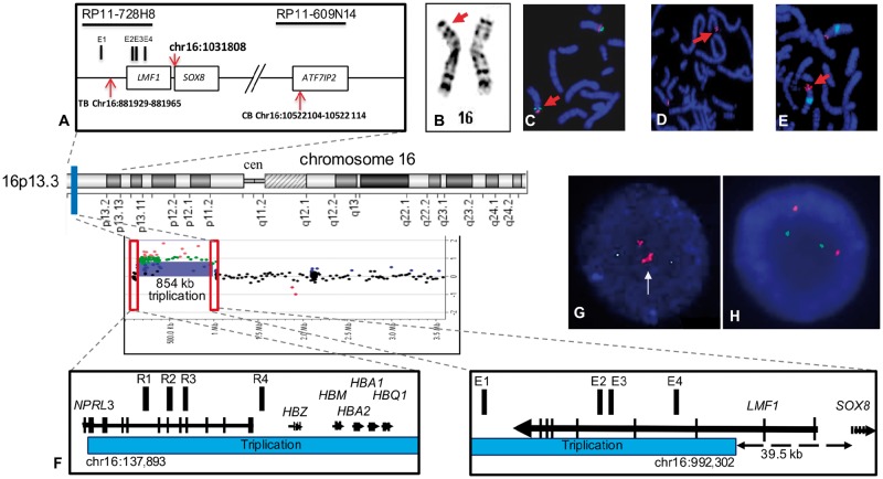
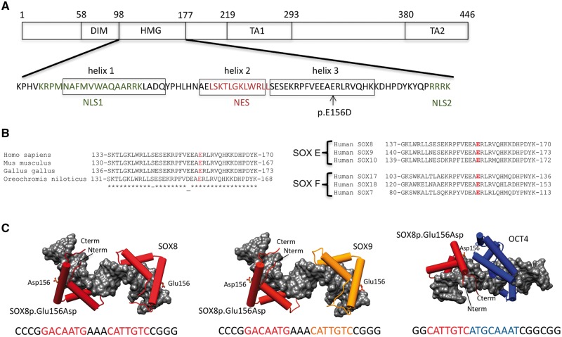
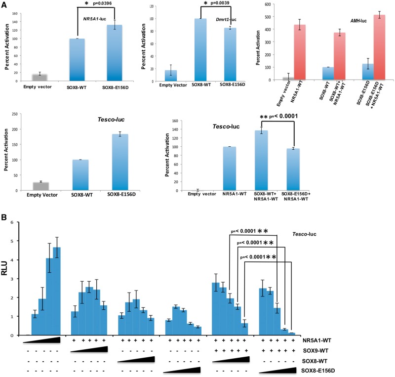
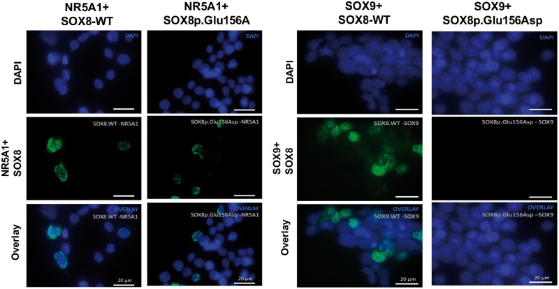
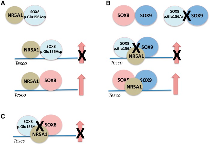
SOX8 is an HMG-box transcription factor closely related to SRY and SOX9. Deletion of the gene encoding Sox8 in mice causes reproductive dysfunction but the role of SOX8 in humans is unknown. Here, we show that SOX8 is expressed in the somatic cells of the early developing gonad in the human and influences human sex determination. We identified two individuals with 46, XY disorders/differences in sex development (DSD) and chromosomal rearrangements encompassing the SOX8 locus and a third individual with 46, XY DSD and a missense mutation in the HMG-box of SOX8. In vitro functional assays indicate that this mutation alters the biological activity of the protein. As an emerging body of evidence suggests that DSDs and infertility can have common etiologies, we also analysed SOX8 in a cohort of infertile men (n = 274) and two independent cohorts of women with primary ovarian insufficiency (POI; n = 153 and n = 104). SOX8 mutations were found at increased frequency in oligozoospermic men (3.5%; P < 0.05) and POI (5.06%; P = 4.5 × 10⁻⁵) as compared with fertile/normospermic control populations (0.74%). The mutant proteins identified altered SOX8 biological activity as compared with the wild-type protein. These data demonstrate that SOX8 plays an important role in human reproduction and SOX8 mutations contribute to a spectrum of phenotypes including 46, XY DSD, male infertility and 46, XX POI.


Figures

Figure 1.
Figure 1.
Chromosomal rearrangements at the SOX8 locus associated with 46, XY DSD. (A) Schematic representation of the 16p inversion in patient 1 indicating the genes and clones of the region and the position of the telomeric breakpoint (TB) and centromeric breakpoints (CB). The position of the transcription initiation site of the SOX8 gene is indicated (chr16: 1 031 808), the Sox8 enhancer elements (E1–E4) and the position of the BACs used for FISH analysis. Patient 1 with (B) GTG banding showing the rearranged chromosome 16 (arrow). (C) FISH on metaphases with BAC RP11–161M6 (green; chr16: 991, 108–1, 141, 991- UCSC Genome Browser; GRCh37/hg19) and RP11–297M9 (red; chr16: 9, 727, 655–9, 920, 250) confirming the paracentric inversion (arrow). (D) The CB is located within the clone, RP11–609N14 on 16p13.13). (E) The TB mapped within RP11–728H8, on 16p13.3 (arrows). Patient 2 (F) Chromosome 16p rearrangement. The aCGH profile indicated an 854 kb triplication in the 16p13.3 region. Affected region is indicated by a blue bar. Red rectangles outline the triplication breakpoints. A magnified view of the triplication breakpoints shows, on the left, a CB located in the intron 1 of the NPRL3 gene. The 16p13.3 triplication starts at chr16: 137893 position and spans proximally including the α-globin genes (HBZ, HBM, HBA1, HBA2, and HBQ1) and their upstream cis-regulatory elements R1-R4 (red bars). On the right, the TB of the triplication (chr16: 992, 302) is located ∼39.5 kb upstream of the SOX8 gene. The SOX8-specific enhancers are mapped within the triplicated segment. Genes are indicated by black arrows with vertical lines specifying exons. Arrows correspond to a direction of gene transcription. (G) Interphase FISH analysis using RP11–598 I20 (16p13.3, labeled red) and a control RP11–121O8 (16p13.1, labeled green) probe showing a triplication (white arrow) in the patient 20. (H) Representative FISH image using the same probes shows a normal hybridization pattern observed in both parents.
Figure 2.
Figure 2.
Gonad histology of Patients 1 and 2. Patient 1, (A) Ovarian-like stromal cells with no evidence of testicular tissue consistent with 46, XY complete gonadal dysgenesis. Patient 2, (B) Seminiferous tubules (cords) are mildly contorted in places (black arrow) and some are misshapen. The interstitium space is expanded and mildly fibrotic (white arrow; H&E; 200X). (C) Immunohistochemistry for calretinin shows only focal Leydig cells (arrows; calretinin stain, blue; 200X). (D) Inhibin immunohistochemistry highlights Sertoli cells, making the contorted tubules more evident (inhibin; 200X). (E) Immunohistochemistry for SOX9 staining the nuclei of Sertoli cells within tortuous tubules (SOX9; 200X).
Figure 3.
Figure 3.
Expression of the SOX8 protein in human gonad tissues. (A) The SOX8 protein (red) is expressed in the human male Sertoli and Leydig cells together with NR5A1 (green) during early testis formation (image at 9 weeks post-conception). Primitive seminiferous cords are indicated by the dashed lines (B) The SOX8 protein expression in the granulosa cells of the late fetal ovary (image at 40 weeks of gestation). (C) Immunohistochemistry showing SOX8 expression in granulosa cells (arrow) lining the follicles of the adult ovary (19 years old; scale bars are 50 μm).
Figure 4.
Figure 4.
Mutation in the HMG-box of SOX8 associated with 46, XY gonadal dysgenesis. (A) Schematic representation showing important functional domains of the SOX8 protein. The amino acid sequence of the HMG domain including the three alpha-helices, the two nuclear localization signals (NLS1, NLS2), and the nuclear export sequence (NES) together with the position of SOX8p.Glu156Asp (p.E156D) mutation are indicated. The DNA-dependent dimerization domain (DIM), the DNA-binding HMG domain, and the transactivation domains (TA1, TA2) are shown. (B) Left, alignment of the C-terminal of the SOX8 HMG-box domain containing helix 3 from selected vertebrates. Right, the alignment of the distal portion of the SOX8 HMG-box domain with other human SOXE and SOXF family members. The position of the p.Glu156Asp missense mutation is highlighted. (C) Structural models for ternary complexes of conjectured SOX8p.Glu156Asp -SOX8, SOX8p.Glu156Asp -SOX9 and SOX8p.Glu156Asp -OCT4 dimers on composite DNA elements. SOX8 is shown in dark red, SOX9 in orange and OCT4 in blue. The HMG domains of SOX E proteins and the POU domain of OCT4 are shown as cartoon with cylindrical alpha-helices and the DNA as gray van-der-Waals surface. The SOX8p.Glu156Asp site is drawn as ball-and-sticks. The DNA sequences used to generate the models with colour-coded binding elements are depicted underneath the models. The SOXE HMG box is N-terminally extended by a 40 amino acid dimerization (DIM) domain of unknown structure that mediates highly cooperative DNA-dependent dimerization presumably by interactions with the HMG box.
Figure 5.
Figure 5.
SOX8p.Glu156Asp shows altered biological activity. (A) The transcriptional activities of WT-SOX8 and SOX8p.Glu156Asp were studied using the human AMH and NR5A1 promoters, mouse Dmrt1 promoter and the Sox9 Tesco enhancer as reporters, following transfection in HEK293-T cells. The data shown represent the mean ± SEM of minimum three independent experiments, each of which was performed at least in quadruplicate. The reporter constructs were transfected into HEK293-T cells with either the WT-SOX8 or SOX8p.Glu156Asp expression vector. The results are expressed as relative percentage of WT-SOX8 activity (100%). The SOX8p.Glu156Asp can activate the gonadal promoters in a similar fashion to WT-SOX8. However, although SOX8p.Glu156Asp can synergistically activate the AMH promoter with NR5A1, it fails to synergise with NR5A1 to activate the Sox9 Tesco enhancer. (B) HEK 293-T cells were co-transfected with Tesco reporter construct (10ng), WT-NR5A1 (1ng) and WT-SOX9 (1ng) and increasing amount of WT-SOX8 or SOX8p.Glu156Asp (0, 1, 2, 5, 10 ng). Results are expressed as Relative Luminescence Units (RLU). The SOX8p.Glu156Asp mutant exerts a repressive effect by preventing synergistic activation of the Sox9 Tesco enhancer by NR5A1 and SOX9 even at three times lower concentrations.
Figure 6.
Figure 6.
Protein–protein interaction assayed using Proximity ligation assay. Plasmids encoding SOX8-WT or SOX8p.Glu156Asp were transiently expressed with WT-NR5A1 or SOX9 for 48 h in HEK293T cells. Protein–protein interaction of SOX8-WT and SOX8p.Glu156Asp with NR5A1 and SOX9 were analysed using the Duolink proximity ligation assay. Nuclei are stained with DAPI (blue) and Duolink signal representing interaction between the proteins is shown in green. Each green dot represents a single dimerization event. Both the SOX8-WT and SOX8p.Glu156Asp proteins can physically interact with NR5A1. Whereas, although SOX8-WT protein can interact with SOX9, the SOX8p.Glu156Asp cannot.
Figure 7.
Figure 7.
Schematic representation of the results of the in vitro assays for the SOX8p.Glu156Asp protein. (A) The mutant SOX8 protein has the ability to physically interact with the NR5A1 protein but in contrast to the WT-SOX8 protein does not synergize with NR5A1 to promote reporter gene activity using the Tesco enhancer as a target. (B) The mutant SOX8 protein does not physically interact with the SOX9 protein and shows a lack of synergy with SOX9/NR5A1 to promote reporter gene activity. (C) The SOX8 mutant protein also shows dominant negative activity by impairing promoter gene activity of the WT-SOX8 protein in synergy with NR5A1.

Similar articles

Cited by

References

    1. Sekido R., Lovell-Badge R. (2008) Sex determination involves synergistic action of SRY and SF1 on a specific Sox9 enhancer. Nature, 453, 930–934. - PubMed
    1. Lin Y.T., Capel B. (2015) Cell fate commitment during mammalian sex determination. Curr. Opin. Genet. Dev., 32, 144–152. - PMC - PubMed
    1. Chaboissier M.C., Kobayashi A., Vidal V.I., Lützkendorf S., van de Kant H.J., Wegner M., de Rooij D.G., Behringer R.R., Schedl A. (2004) Functional analysis of Sox8 and Sox9 during sex determination in the mouse. Development, 131, 1891–1901. - PubMed
    1. Lavery R., Chassot A.A., Pauper E., Gregoire E.P., Klopfenstein M., de Rooij D.G., Mark M., Schedl A., Ghyselinck N.B., Chaboissier M.C. (2012) Testicular differentiation occurs in the absence of R-spondin1 and Sox9 in mouse sex reversals. PLoS Genet., 8, e1003170.. - PMC - PubMed
    1. Barrionuevo F., Georg I., Scherthan H., Lécureuil C., Guillou F., Wegner M., Scherer G. (2009) Testis cord differentiation after the sex determination stage is independent of Sox9 but fails in the combined absence of Sox9 and Sox8. Dev. Biol., 327, 301–312. - PubMed

Publication types

Substances