. 2018 Jan 25;172(3):590-604.e13.
doi: 10.1016/j.cell.2017.12.032.

Context-Dependent and Disease-Specific Diversity in Protein Interactions within Stress Granules

Affiliations

Context-Dependent and Disease-Specific Diversity in Protein Interactions within Stress Granules

Sebastian Markmiller et al. Cell. .

Abstract

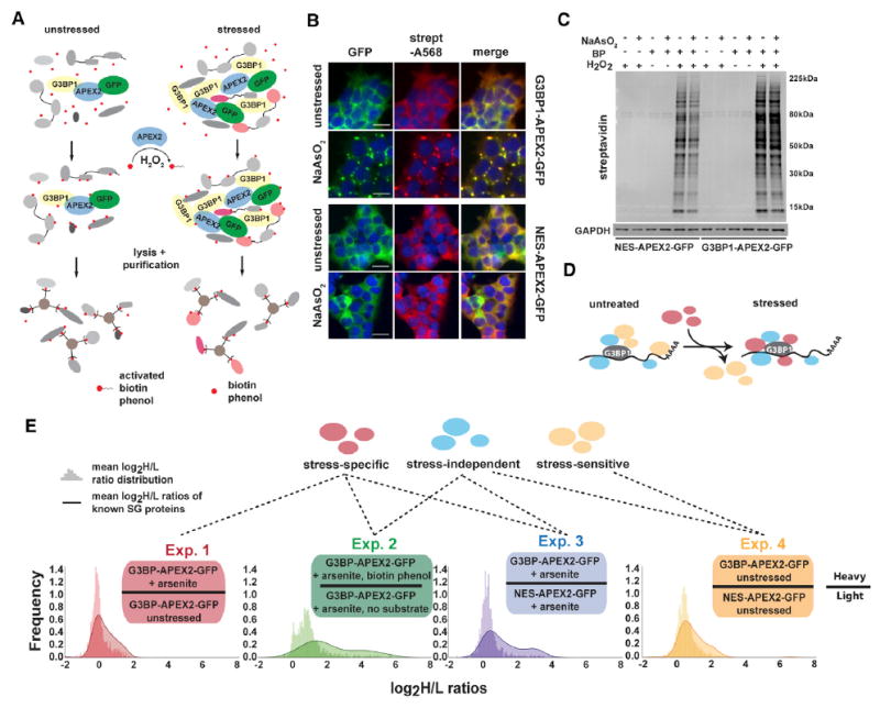
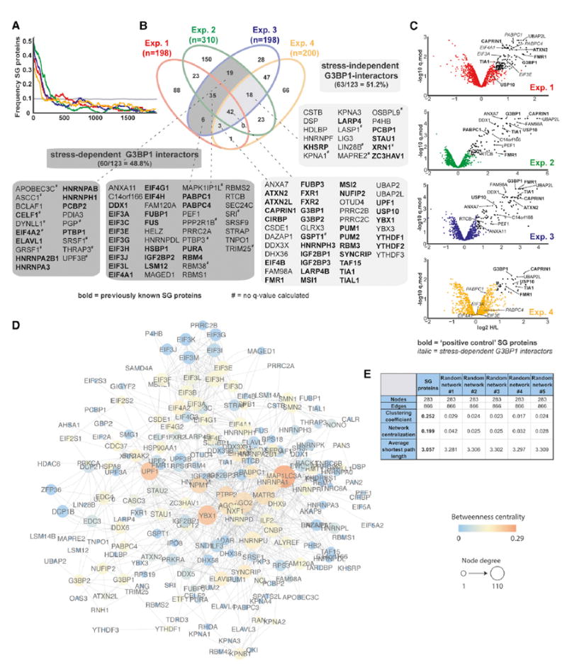
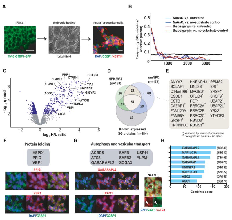
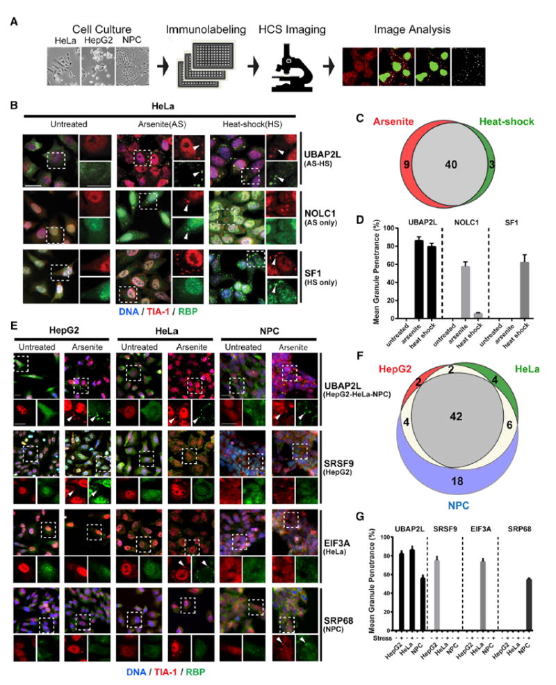
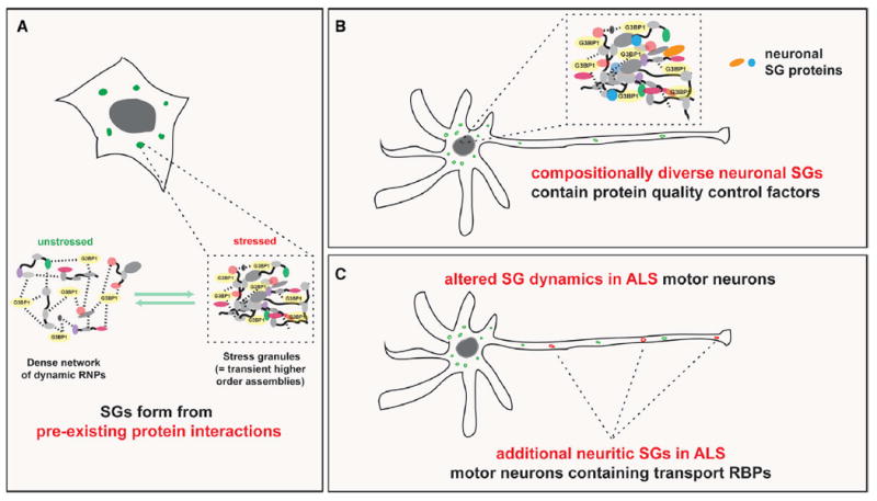
Stress granules (SGs) are transient ribonucleoprotein (RNP) aggregates that form during cellular stress and are increasingly implicated in human neurodegeneration. To study the proteome and compositional diversity of SGs in different cell types and in the context of neurodegeneration-linked mutations, we used ascorbate peroxidase (APEX) proximity labeling, mass spectrometry, and immunofluorescence to identify ∼150 previously unknown human SG components. A highly integrated, pre-existing SG protein interaction network in unstressed cells facilitates rapid coalescence into larger SGs. Approximately 20% of SG diversity is stress or cell-type dependent, with neuronal SGs displaying a particularly complex repertoire of proteins enriched in chaperones and autophagy factors. Strengthening the link between SGs and neurodegeneration, we demonstrate aberrant dynamics, composition, and subcellular distribution of SGs in cells from amyotrophic lateral sclerosis (ALS) patients. Using three Drosophila ALS/FTD models, we identify SG-associated modifiers of neurotoxicity in vivo. Altogether, our results highlight SG proteins as central to understanding and ultimately targeting neurodegeneration.

Keywords: RNA-binding proteins; amyotrophic lateral sclerosis; granules; heat shock; motor neuron disease; neurodegeneration; phase separation; ribonucleoprotein; stress.


Conflict of interest statement

DECLARATION OF INTERESTS

G.W.Y. is a co-founder of Locana and Eclipse Bioinnovations and member of the scientific advisory boards of Locana, Eclipse Bioinnovations and Aquinnah Pharmaceuticals. The terms of this arrangement have been reviewed and approved by the University of California, San Diego in accordance with its conflict of interest policies. A.S. and M.W.K. are employees of Biogen. All other authors declare no competing interests

Figures

Figure 1
Figure 1. G3BP1-APEX2 Mediates Specific Biotinylation of Stress-Granule-Associated Proteins
(A) Schematic of APEX proximity labeling to tag SG proteins with biotin. (B) Streptavidin staining of unstressed and NaAsO2-treated HEK293T G3BP1-APEX2-GFP and hPGK-NES-APEX2-GFP cells. Scale bars, 25 μm. (C) Streptavidin-HRP western blot analysis of induced protein biotinylation in lysates from NES-APEX2-GFP and G3BP1-APEX2-GFP cells. (D) Schematic of G3BP1 interactome changes upon stress. (E) Experimental designs for detecting the G3BP1 interactome changes under different conditions, including log2 H/L ratio distributions of all proteins detected, overlaid with log2 H/L ratio distributions of known SG proteins. See also Figures S1 and S2 and Table S1.
Figure 2
Figure 2. SG-APEX Identifies Known and Previously Unknown SG Proteins within a Dense Interaction Network
(A) Enrichment frequency distribution of known SG proteins in log2 H/L-ranked proteomics datasets. The dashed line represents 2 times the background frequency of SG proteins across all detected proteins. (B) Venn diagram showing overlapping hits from four experimental designs, with previously known SG proteins highlighted in bold. (C) Volcano plots showing statistically significant enrichment of selected known and previously unknown SG proteins across experiments. (D) Protein interaction network (PIN) of 283 proteins identified as APEX hits in HEK293T cells or previously shown to associate with SGs. Network was visualized in Cytoscape using a force-directed layout. (E) Common network parameters for the SG-PIN compared to five PINs from a randomly selected equal number of nodes and edges. See also Table S3.
Figure 3
Figure 3. NPCs and HEK293T Cells Contain Distinct but Overlapping Sets of SG Proteins
(A) Overview of NPC generation from induced pluripotent stem cells (iPSCs). (B) Enrichment frequency distribution of known SG proteins in log2 H/L-ranked proteomics datasets. The dashed lines represent 2 times the background frequency of SG proteins across all detected proteins. (C) Volcano plot showing statistically significant enrichment of selected known and previously unknown neuronal SG proteins in NPCs. (D) Venn diagram showing the overlap between known SG proteins and SG-APEX hits identified in HEK293T cells and NPCs. (E) Previously unknown SG proteins identified by SG-APEX in both HEK293T cells and NPCs. (F) IF images of selected neuronal SG proteins with functions related to protein folding. (G) IF images of selected neuronal SG proteins with functions in autophagy and vesicular transport. (H) Ranked list of proteins with the greatest connectivity to SG proteins as determined by the Enrichr gene enrichment analysis tool. Scale bars in (A), (F), and (G), 25 μm. See also Figure S3 and Table S4.
Figure 4
Figure 4. An RBP-Centered Imaging Screen Identifies Stress- and Cell-Type-Specific SG Components
(A) High-content imaging (HCI) screen outline to identify SG-localized RBPs in HepG2 cells, HeLa cells, and NPCs. (B) IF images showing examples of RBP localization in untreated, NaAsO2 (AS)-treated, and heat-shock (HS)-treated HeLa cells. UBAP2L is a common hit in both stress conditions; NOLC1 and SF1 are specific to NaAsO2 and heat shock, respectively. Left panels are merged lower-resolution views, and right panels represent zoomed-in views of the indicated selection separately showing TIA-1 (red) or the test RBP (green). Arrowheads indicate co-localization of the test RBP with TIA1. (C) Venn diagram comparing SG proteins in HeLa cells treated with NaAsO2 versus heat shock. (D) Quantification of the mean granule penetrance of proteins with either constitutive (UBAP2L) or stress-type-specific (NOLC1 and SF1) SG localization. (E) IF images showing examples of RBP localization in untreated and NaAsO2-treated HeLa cells, HepG2 cells, or NPCs. UBAP2L is found in SGs in all three cell types, while SRSF9, EIF3A, and SRP68 are specific to HepG2 cells, HeLa cells, and NPCs, respectively. Top panels are merged lower-resolution views, while the bottom panels represent zoomed-in views of the indicated selection separately showing TIA-1 (red) or the test RBP (green). Arrowheads indicate examples of RBPs co-localized with TIA-1. (F) Venn diagram comparing SG proteins in HepG2, HeLa and NPCs treated with NaAsO2. (G) Mean granule penetrance of proteins with either cell-type-independent or cell-type-specific SG localization. Scale bars in (B) and (E), 20 μm. Error bars in (D) and (G) represent SD. See also Table S5.
Figure 5
Figure 5. SG Composition and Subcellular Distribution Is Altered in ALS-Patient-Derived iPS-MNs
(A) IF images of SND1 and IGF2BP3 localization in unstressed or NaAsO2-treated iPS-MNs. Top panels are merged lower-resolution views, while the bottom panels represent zoomed-in views of the indicated selection separately showing G3BP1 (green) or the test RBP (red). Arrowheads indicate examples of RBPs co-localized with G3BP1. (B) Overview of RBPs whose localization in unstressed iPS-MNs is either restricted to the cell body or extends into neuronal projections. (C) Time-course analysis of SG formation in iPS-MNs from controls or from ALS patients bearing mutations in HNRNPA2B1 or C9orf72, respectively. (D) IF images of control and HNRNPA2B1 mutant iPS-MNs that were either untreated or stressed with puromycin. Top panels are merged lower-resolution views, while the bottom panels represent zoomed-in views of the indicated selection separately showing G3BP1 (green) or the test RBP (red). White and yellow arrowheads indicate examples of SGs formed in cell bodies or neurites, respectively. (E) Quantification of SG area and number in untreated or stressed control and HNRNPA2B1 mutant iPS-MNs. (F) Quantification of RBPs that localize to SGs in cell bodies or dendritic projections in control versus HNRNPA2B1 mutant cells. The RBPs exhibiting targeting to SGs in projections in HNRNPA2B1 mutant cells are highlighted in the panel on the right. Scale bars, 20 μm. Error bars in (C) and (E) represent SD. Statistical significance was calculated by 2-way ANOVA (C) or Student’s t test (E). *p ≤ 0.05; **p ≤ 0.01; ***p ≤ 0.001; ****p ≤ 0.0001. See also Figure S4 and Table S6.
Figure 6
Figure 6. Integrative Data Analysis Highlights Potential Disease-Relevant Proteins
(A) Venn diagram showing overlap between proteins identified in our combined APEX-IF approach, known SG proteins, and RBPs. (B) Protein domain enrichment analysis of 260 SG APEX-IF. (C) Gene ontology analysis for 260 APEX-IF hits. (D) Comparison of the proportion of amino acids in LCDs and IDRs between APEX-IF hits and background. (E) Heatmap for the 75 proteins most broadly represented across selected SG and neurodegeneration-relevant datasets. Heatmap indicates whether a protein is present (blue box) or absent (white box) from each dataset, and proteins are ranked by the number of datasets they are part of in descending order from left to right. (F) Images of Drosophila eye degeneration models crossed with the indicated strains. (G) Images and quantitation of the wing notching phenotype caused by poly(GR) toxicity in flies. w1118 flies were used as the control for genetic mutant alleles, while UAS-GFP served as the control for different UAS-RNAi lines. Numbers indicate Bloomington stock numbers for each mutant or RNAi line. (H) IF images of G3BP1 staining and quantification of SG numbers in HeLa cells treated with control siRNA or siRNA targeting UBAP2L. Data are presented as mean ± SEM, and statistical significance was determined by two-tailed unpaired t test. (I) IF images of 293FITR cells with inducible expression of either a full-length UBAP2L-mCherry fusion protein (top panel) or a truncated UBAP2L-mCherry fusion protein missing the N-terminal UBA domain (middle and bottom panels). Scale bars in (H) are 25 μm. See also Figure S5 and Table S7.
Figure 7
Figure 7. SG Form from Pre-existing PPIs Are Especially Diverse in Neuronal Cells and Display Aberrant Characteristics in ALS Mutant Cells
(A) Model of the relationships among normally functioning, dynamic RNPs, transient SGs, and permanent pathological protein inclusions. (B) Schematic showing that neuronal SGs are diverse and contain proteins involved in protein quality control pathways. (C) Schematic showing altered SG dynamics, composition, and subcellular distribution in ALS mutant motor neurons.

Comment in

References

    1. Ainger K, Avossa D, Morgan F, Hill SJ, Barry C, Barbarese E, Carson JH. Transport and localization of exogenous myelin basic protein mRNA microinjected into oligodendrocytes. J Cell Biol. 1993;123:431–441. - PMC - PubMed
    1. Alami NH, Smith RB, Carrasco MA, Williams LA, Winborn CS, Han SSW, Kiskinis E, Winborn B, Freibaum BD, Kanagaraj A, et al. Axonal transport of TDP-43 mRNA granules is impaired by ALS-causing mutations. Neuron. 2014;81:536–543. - PMC - PubMed
    1. Aulas A, Fay MM, Lyons SM, Achorn CA, Kedersha N, Anderson P, Ivanov P. Stress-specific differences in assembly and composition of stress granules and related foci. J Cell Sci. 2017;130:927–937. - PMC - PubMed
    1. Becker LA, Huang B, Bieri G, Ma R, Knowles DA, Jafar-Nejad P, Messing J, Kim HJ, Soriano A, Auburger G, et al. Therapeutic reduction of ataxin-2 extends lifespan and reduces pathology in TDP-43 mice. Nature. 2017;544:367–371. - PMC - PubMed
    1. Beckmann BM, Horos R, Fischer B, Castello A, Eichelbaum K, Alleaume AM, Schwarzl T, Curk T, Foehr S, Huber W, et al. The RNA-binding proteomes from yeast to man harbour conserved enigmRBPs. Nat Commun. 2015;6:10127. - PMC - PubMed

Publication types

Substances

LinkOut - more resources