Differential Response of B Cells to an Immunogen, a Mitogen and a Chemical Carcinogen in a Mouse Model System
- PMID: 29373896
- PMCID: PMC5844640
- DOI: 10.22034/APJCP.2018.19.1.81
Differential Response of B Cells to an Immunogen, a Mitogen and a Chemical Carcinogen in a Mouse Model System
Abstract
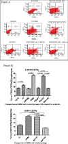
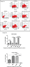
Background: B cells are specific antibody generating cells which respond to foreign intruders in the circulation. The purpose of this study was to compare the relative immunogenic potentials of three well established agent types viz. an immunogen, a mitogen and a carcinogen, by following B cell responses to their presence in a mouse model system. Methods: Mice were treated with tetanus toxoid (immunogen), poke weed mitogen (typical mitogen), and benzo-α- pyrene (carcinogen) and generated B cell populations were determined in isolated splenic lymphocytes (splenocytes) by flow cytometry using specific anti-B cell marker antibodies. Flow cytometric estimation of LDL receptor (LDLR) expression, along with associated B cell markers, was also conducted. Kit based estimation of serum IgG, western blotting for LDLR estimation on total splenocytes and spectrometry for cholesterol and serum protein estimation were further undertaken. Student’s T-tests and one way ANOVA followed by the Bonferroni method were employed for statistical analysis. Results: The mitogen was found to better stimulate B cell marker expression than the immunogen, although the latter was more effective at inducing antibody production. The chemical carcinogen benzo-α-pyrene at low concentration acted potentially like a mitogen but almost zero immunity was apparent at a carcinogenic dose, with a low profile for LDLR expression and intracellular cholesterol. Conclusion: The findings in our study demonstrate an impact of concentration of BaP on generation of humoral immunity. Probably by immunosuppression through restriction of B-cell populations and associated antibodies, benzo-α-pyrene may exerts carcinogenicity. The level of cholesterol was found to be a pivotal target.
Keywords: B cells; IgG; LDL receptor; cholesterol; tetanus toxoid; pokeweed mitogen; benzo; alpha; pyrene.
Creative Commons Attribution License
Figures


Similar articles
-
Differential response of T cells to an immunogen, a mitogen and a chemical carcinogen in a mouse model system.J Biochem Mol Toxicol. 2019 May;33(5):e22290. doi: 10.1002/jbt.22290. Epub 2019 Jan 21. J Biochem Mol Toxicol. 2019. PMID: 30664314
-
Cholesterol homeostasis and cell proliferation by mitogenic homologs: insulin, benzo-α-pyrene and UV radiation.Cell Biol Toxicol. 2018 Aug;34(4):305-319. doi: 10.1007/s10565-017-9415-8. Epub 2017 Nov 3. Cell Biol Toxicol. 2018. PMID: 29101605
-
Evaluation of murine splenic cell type metabolism of benzo[a]pyrene and functionality in vitro following repeated in vivo exposure to benzo[a]pyrene.Toxicol Appl Pharmacol. 1992 Oct;116(2):258-66. doi: 10.1016/0041-008x(92)90305-c. Toxicol Appl Pharmacol. 1992. PMID: 1412470
-
Direct suppression of in vitro antibody production by mouse spleen cells by the carcinogen benzo(a)pyrene but not by the noncarcinogenic congener benzo(e)pyrene.Cancer Res. 1984 Aug;44(8):3388-93. Cancer Res. 1984. PMID: 6331644
-
Genetic immunization converts the trypanosoma cruzi B-Cell mitogen proline racemase to an effective immunogen.Infect Immun. 2010 Feb;78(2):810-22. doi: 10.1128/IAI.00926-09. Epub 2009 Nov 16. Infect Immun. 2010. PMID: 19917711 Free PMC article.
References
-
- Albi E, Cataldi S, Rossi G, Magni MV. A possible role of cholesterol-sphingomyelin/phosphatidylcholine in nuclear matrix during rat liver regeneration. J Hepatol. 2003;38:623–8. - PubMed
-
- Albi E, Magni M. The role of intranuclear lipids. Biol Cell. 2004;96:657–7. - PubMed
-
- Albi E. Role of intranuclear lipids in health and disease. Clin Lipidol. 2011;6:59–69.
-
- Balakrishnan K, Adams LE. The role of the lymphocyte in an immune response. Immunol Invest. 1995;24:233–4. - PubMed

