. 2018 Mar 1;200(5):1817-1828.
doi: 10.4049/jimmunol.1700639. Epub 2018 Jan 26.

PAD4 Deficiency Leads to Decreased Organ Dysfunction and Improved Survival in a Dual Insult Model of Hemorrhagic Shock and Sepsis

Affiliations

PAD4 Deficiency Leads to Decreased Organ Dysfunction and Improved Survival in a Dual Insult Model of Hemorrhagic Shock and Sepsis

Bethany M Biron et al. J Immunol. .

Abstract

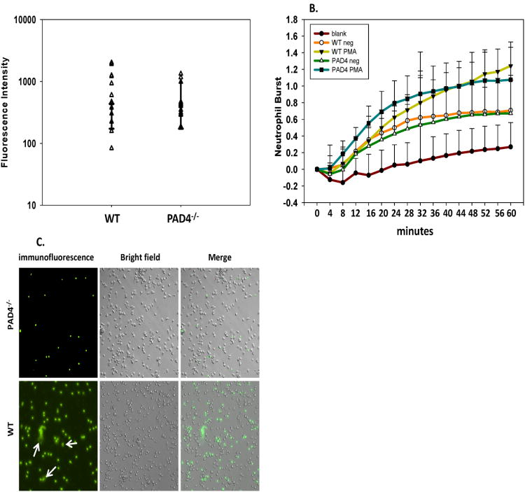
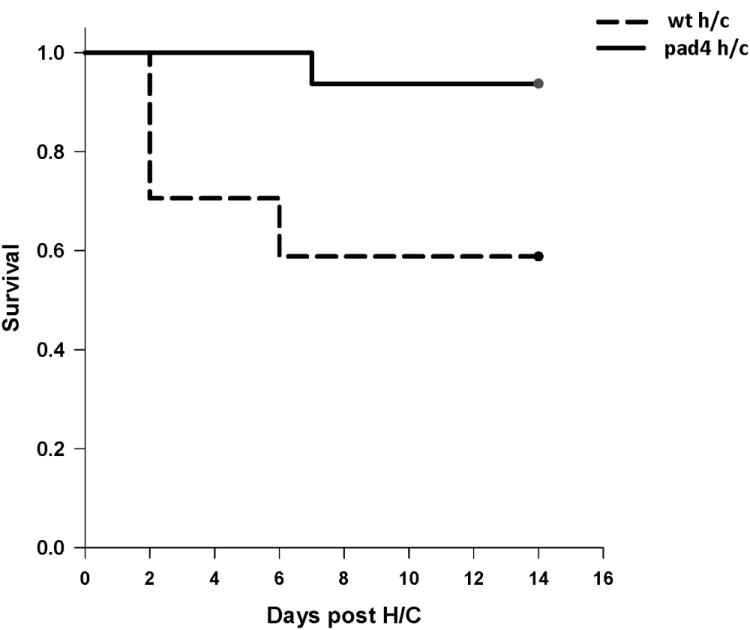
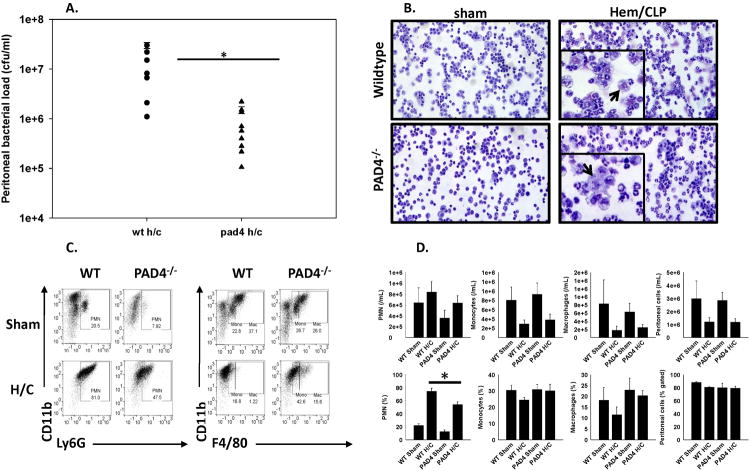
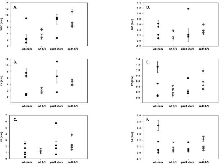
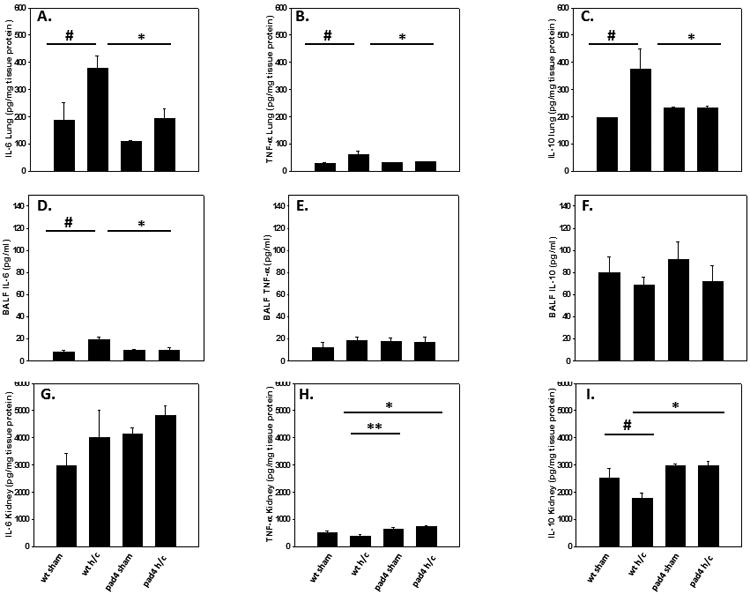
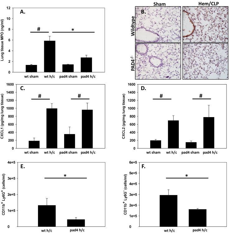
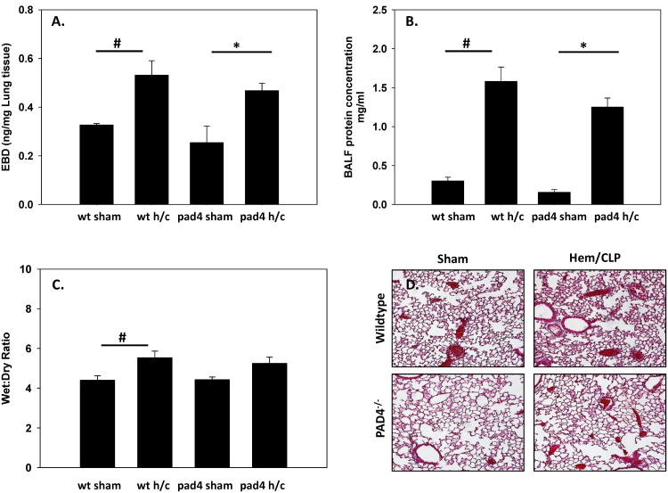
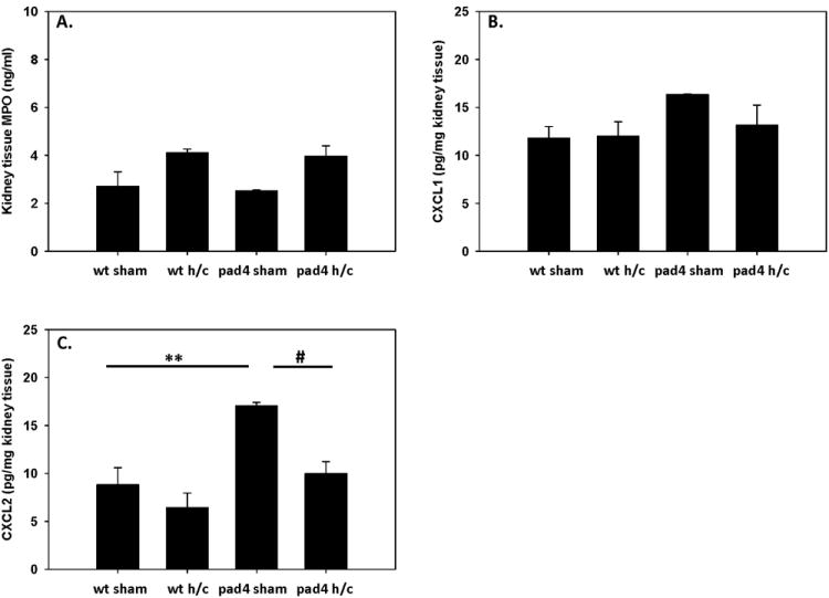
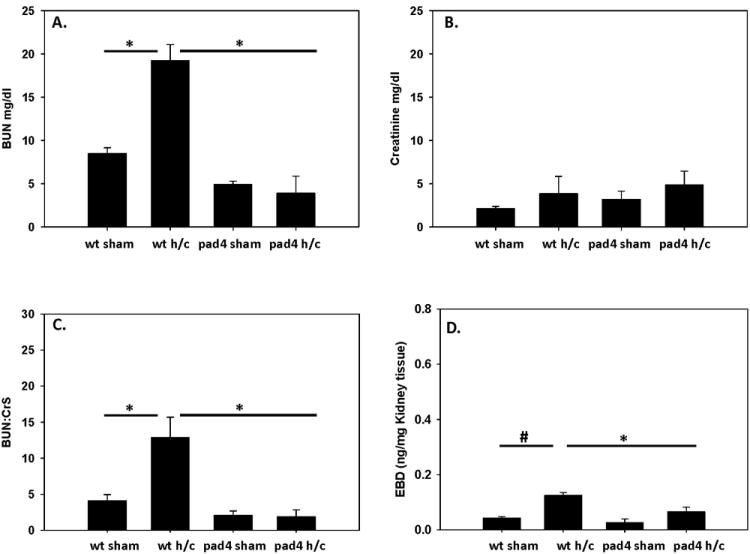
Indirect acute respiratory distress syndrome (iARDS) is caused by a nonpulmonary inflammatory process resulting from insults such as nonpulmonary sepsis. Neutrophils are thought to play a significant role in mediating ARDS, with the development of iARDS being characterized by dysregulation and recruitment of activated neutrophils into the lung. Recently, a novel mechanism of microbial killing by neutrophils was identified through the formation of neutrophil extracellular traps (NETs). NETs are composed of large webs of decondensed chromatin released from activated neutrophils into the extracellular space; they are regulated by the enzyme peptidylarginine deiminase 4 (PAD4) through mediation of chromatin decondensation via citrullination of target histones. Components of NETs have been implicated in ARDS. However, it is unknown whether there is any pathological significance of NET formation in ARDS caused indirectly by nonpulmonary insult. We subjected PAD4-/- mice and wild-type mice to a "two-hit" model of hypovolemic shock (fixed-pressure hemorrhage [Hem]) followed by septic cecal ligation and puncture (CLP) insult (Hem/CLP). Mice were hemorrhaged and resuscitated; 24 h after Hem, mice were then subjected to CLP. Overall, PAD4 deletion led to an improved survival as compared with wild-type mice. PAD4-/- mice displayed a marked decrease in neutrophil influx into the lung, as well decreased presence of proinflammatory mediators. PAD4-/- mice were also able to maintain baseline kidney function after Hem/CLP. These data taken together suggest PAD4-mediated NET formation contributes to the mortality associated with shock/sepsis and may play a role in the pathobiology of end organ injury in response to combined hemorrhage plus sepsis.


Figures

Figure 1
Figure 1. PAD4 gene deficiency effects NET formation but does not inhibit other neutrophil effector functions
Neutrophils from naïve C57BL/6 (WT) and PAD4-/- mice were analyzed for other anti-microbial functions ex-vivo. Phagocytosis capabilities were comparable in WT and PAD4-/- mice (A). ROS generation after stimulation with PMA was increased in WT and PAD4-/- mice (B). Neutrophils were isolated via thioglycolate injection and were cultured on Poly-L-lysine coated plates for 4 hours post 20nM PMA stimulation to elicit NET production. NETs were visualized via immunofluorescence with Sytox green (C). PAD4-/- neutrophils displayed a deficiency in producing NETs, while WT neutrophils were able to produce NET structures after PMA stimulation. n=3 independent experiments/samples run in triplicate
Figure 2
Figure 2. PAD4-deficient mice display improved survival in Hem/CLP model of iARDS
Mice were subjected to hemorrhage and subsequent septic insult and then monitored daily for 14 days. PAD4-/- mice had 90% survival at 14 days compared to the 50% survival in WT mice. Sham animals from both groups had a 100% survival rate. log-rank test. p=0.013. (n=6 sham/sham (not shown); n=16-17 Hem/CLP per group)
Figure 3
Figure 3. PAD4-/- mice exhibit enhanced bacterial clearance at site of infection after Hem/CLP
Twenty four hours after Hem/CLP, peritoneal cells were collected and assessed for neutrophil/macrophage recruitment as well as antimicrobial function. Lavage contents collected from the peritoneum 24 hrs post Hem/CLP were placed on blood agar plates for 24 hrs at 37°C. PAD4-/- mice had significantly reduced colony formations compared to WT mice. Sham animals from both groups had no colony formations (A). Cytospins of peritoneal cells were visualized with Giemsa 20× using RGB, DIC N1 filter (0.33μM/pixel). Morphological differences were noted between macrophage populations with WT Hem/CLP peritoneal macrophages appearing more granulated compared to the peritoneal macrophages from the PAD4-/- Hem/CLP mice. Cell morphology was similar in both sham groups (B). The abundance of different leukocyte populations in the peritoneum was measured by flow cytometry based on Ly6G, CD11b, and F4/80 staining (C-D). In absolute number, there was no difference in neutrophils, monocytes, and macrophages between WT and PAD4-/- mice after Hem/CLP. The percentage of neutrophils was decreased in PAD4-/- mice, and, though no significant difference was found, there seems to be a trend toward increased frequency of monocytes and macrophages in PAD4-/- mice after Hem/CLP. One way ANOVA, #p≤0.05 sham vs. Hem/CLP; * p≤0.05 WT Hem/CLP vs PAD4-/- Hem/CLP. n=7-8/group
Figure 4
Figure 4. Circulating neutrophil counts are equivalent between PAD4-/- and WT mice after Hem/CLP
Whole blood from WT and PAD4-/- mice was analyzed for circulating leukocyte cell totals after Hem/CLP. Total white blood count (WBC) (A), lymphocytes (B), neutrophils (C), monocytes (D), eosinophils (E), and basophils (F) were analyzed. Overall, there was a great deal of variability in WT and PAD4-/- sham groups. While leukocyte counts were more consistent after Hem/CLP, numbers were comparable between both groups with no significant differences noticed. n=6-9/group.
Figure 5
Figure 5. PAD4-/- results in decreased pro-inflammatory and anti-inflammatory cytokine levels in the lungs of Hem/CLP mice
Pro-inflammatory and anti-inflammatory cytokine levels were assessed in lung tissue, the bronchial-aveolar lavage fluid (BALF) and kidney tissue of WT and PAD4-/- mice. Within the lung tissue, there were significant decreases in IL-6 (A), TNF-α (B) and IL-10 levels (C) in PAD4-/- after Hem/CLP. In the BALF, PAD4-/- displayed no increase in IL-6 levels, unlike the WT mice (D), with no differences detected in TNF-α (E) and IL-10 levels (F) between groups. In the kidney, there were no differences in IL-6 levels (G). PAD4-/- kidney TNF-α levels were higher compared to WT after hem/CLP (H), however, TNF-α levels between PAD4-/- sham and PAD4-/- Hem/CLP was unchanged. IL-10 levels in WT mice were decreased in the kidney compared to WT sham levels after Hem/CLP (I), while In PAD4-/-, IL-10 levels in the kidney were unchanged between sham and Hem/CLP animals. One Way ANOVA, #p≤0.05 sham vs. Hem/CLP; * p≤0.05 WT Hem/CLP vs PAD4-/- Hem/CLP; ** p≤0.05 WT Hem/CLP vs PAD4-/- sham. Lung tissue n=5-7/group, BALF n=6-9/group, Kidney tissue n=5-9/group
Figure 6
Figure 6. Neutrophil influx and MPO activity is decreased in PAD4-/- mouse lung tissue following Hem/CLP
Lung tissue was assessed for neutrophil infiltration and activation. MPO levels were significantly decreased in PAD4-/- compared to WT mice after Hem/CLP when analyzed via ELISA (A) as well as histologically (B). Chemoattractants CXCL-1 (C) and CXCL-2 (D) were up-regulated in homogenized WT and PAD4-/- lung tissue after Hem/CLP. While no difference in chemoattractant levels, overall neutrophils (as defined as Ly6G+ CD11B+ cells) were significantly decreased in PAD4-/- lung homogenates after Hem/CLP via flow cytometry (E) and also displayed a significant decrease in Ly6G- CD11B+ cells (F). One way ANOVA, #p≤0.05 sham vs. Hem/CLP; * p≤0.05 WT Hem/CLP vs PAD4-/- Hem/CLP. n=6-9/group
Figure 7
Figure 7. Indices of Lung Injury after Hem/CLP
Different parameters of lung injury were assessed in both WT and PAD4-/- after Hem/CLP to gauge lung pathology. Vascular permeability was assessed using Evans Blue Dye extravasation assay 24 hrs after Hem/CLP. No difference in vascular leakage between WT and PAD4-/- mice was detected after Hem/CLP, with both groups exhibiting a similar rise in lung permeability (A). This was consistent with an increase in protein concentrations seen in the BALF in WT and PAD4-/- mice (B). Lung wet:dry ratios showed an increase in lung edema compared to sham mice in both groups (C.) Histological analysis of the left lung lobe via H&E staining displays an increase in interstitial thickening and alveolar congestion in the WT lung after Hem/CLP. This is mitigated in the PAD4-/- lung (D). One Way ANOVA, #p≤0.05 sham vs. Hem/CLP; * p≤0.05 WT Hem/CLP vs PAD4-/- Hem/CLP. Lung samples n=3-8/group, BALF n=3-6/group
Figure 8
Figure 8. MPO levels are not altered in the kidneys of WT compared PAD4-/- mice following Hem/CLP
Homogenized kidney tissue was assessed for MPO activity after Hem/CLP. There were no significant changes detected in MPO levels between WT and PAD4-/- groups (A). CXCL-1 levels were unchanged in the kidney across all groups (B), while CXCL-2 levels were significantly higher in the sham PAD4-/- kidneys as compared to WT (C). However, after Hem/CLP, CXCL2 levels in the PAD4-/- were decreased to comparable WT levels. One way ANOVA, #p≤0.05 sham vs. Hem/CLP; **p≤0.05 WT sham vs PAD4-/- sham. n=6-9/group
Figure 9
Figure 9. PAD4-/- mice maintain kidney function after Hem/CLP
Indices of kidney function was measured in WT and PAD4-/- mice after Hem/CLP. BUN and Creatinine levels were measured in plasma as a measurement of renal function. BUN levels were comparable to sham levels in the PAD4-/- mice whereas WT had significantly increased levels after Hem/CLP (A) while creatinine levels in both groups were similar to their correspondent sham levels (B). When calculating BUN:Cr ratio WT mice displayed a significant increase in BUN:Cr ratios, whereas PAD4-/- remained at sham levels (C). Vascular permeability was assessed using Evans Blue Dye extravasation assay 24 hrs after Hem/CLP. Kidney permeability was significantly decreased in the PAD4-/- after Hem/CLP as measured by a significant decline of Evans Blue Dye recovered from the kidneys (D). One Way ANOVA, #p≤0.05 sham vs. Hem/CLP; * p≤0.05 WT Hem/CLP vs PAD4-/- Hem/CLP. n=6-9/group BUN and Cr assays n=3-6/group EBD assay.

References

    1. Lomas-Neira J, Chung CS, Perl M, Gregory S, Biffl W, Ayala A. Role of alveolar macrophage and migrating neutrophils in hemorrhage-induced priming for ALI subsequent to septic challenge. Am J Physiol Lung Cell Mol Physiol. 2006;290:L51–8. - PubMed
    1. Perl M, Lomas-Neira J, Venet F, Chung CS, Ayala A. Pathogenesis of indirect (secondary) acute lung injury. Expert Rev Respir Med. 2011;5:115–26. - PMC - PubMed
    1. Venet F, Chung CS, Huang X, Lomas-Neira J, Chen Y, Ayala A. Lymphocytes in the development of lung inflammation: a role for regulatory CD4+ T cells in indirect pulmonary lung injury. J Immunol. 2009;183:3472–80. - PMC - PubMed
    1. Wheeler AP, Bernard GR. Acute lung injury and the acute respiratory distress syndrome: a clinical review. Lancet. 2007;369:1553–64. - PubMed
    1. Rubenfeld GD, Caldwell E, Peabody E, Weaver J, Martin DP, Neff M, Stern EJ, Hudson LD. Incidence and outcomes of acute lung injury. N Engl J Med. 2005;353:1685–93. - PubMed

Publication types

MeSH terms