

Endoscopic non-ablative fractional laser therapy in an orthotopic colon tumour model
- PMID: 29374265
- PMCID: PMC5785993
- DOI: 10.1038/s41598-018-19792-2
Endoscopic non-ablative fractional laser therapy in an orthotopic colon tumour model
Abstract
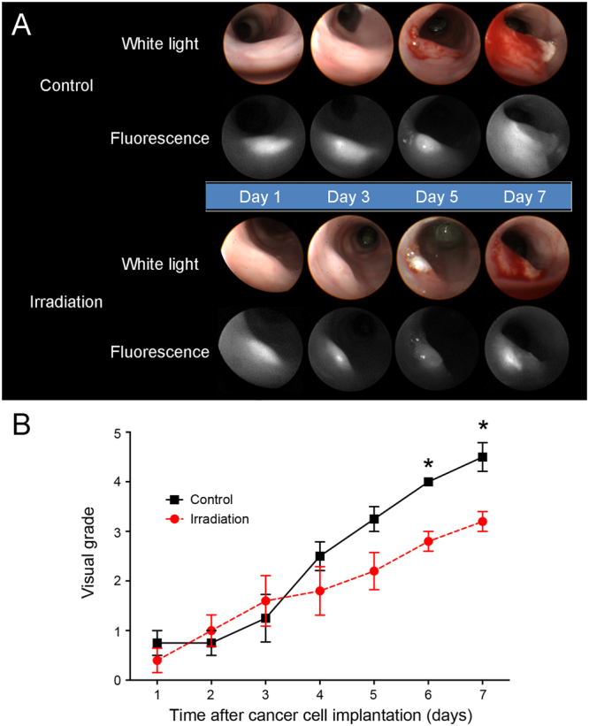
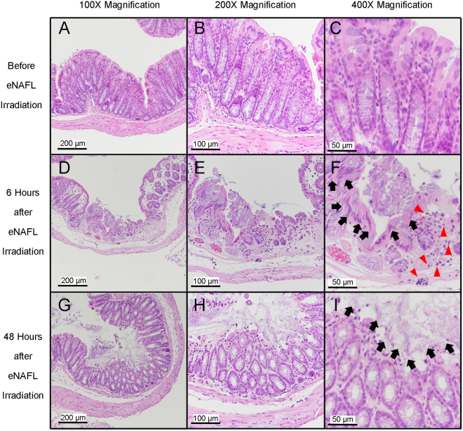
Colorectal cancer is one of the leading causes of cancer-related deaths. Although several therapeutic management strategies are available at the early colon cancer stages, such as endoscopic mucosal or submucosal dissection, associated complications often include bleeding or bowel perforations. As an alternative approach, we investigated endoscopic non-ablative fractional laser (eNAFL) irradiation as a minimally invasive therapeutic modality for the treatment of early-stage colorectal cancer. By implanting SL4-DsRed colon cancer cells into the colons of the C57BL/6 mice, we developed an orthotopic colon tumour mouse model and demonstrated the early-stage tumour growth delay following the eNAFL irradiation. Additionally, we evaluated the temperature changes in the eNAFL-irradiated area using numerical simulations, and induced inflammation using histological analysis. Our results indicate a minimal thermal damage confined to the irradiated spot, sparing the adjacent tissue and alteration in the tumour microenvironment. eNAFL irradiation may be clinically useful as a minimally invasive therapeutic intervention at the early stage of tumourigenesis. In future, an optimal eNAFL therapeutic dose should be determined, in order to increase the efficacy of this approach.
Conflict of interest statement
The authors declare that they have no competing interests.
Figures


Similar articles
-
Effects of deviation from focal plane on lesion geometry for ablative fractional photothermolysis.Lasers Surg Med. 2016 Jul;48(5):555-61. doi: 10.1002/lsm.22481. Epub 2016 Feb 4. Lasers Surg Med. 2016. PMID: 26842919
-
Correlation of histological findings of single session Er:YAG skin fractional resurfacing with various passes and energies and the possible clinical implications.Lasers Surg Med. 2008 Mar;40(3):171-7. doi: 10.1002/lsm.20607. Lasers Surg Med. 2008. PMID: 18366083 Clinical Trial.
-
Animal models of human colorectal cancer: Current status, uses and limitations.World J Gastroenterol. 2015 Nov 7;21(41):11854-61. doi: 10.3748/wjg.v21.i41.11854. World J Gastroenterol. 2015. PMID: 26557009 Free PMC article.
-
Defeating Cancers' Adaptive Defensive Strategies Using Thermal Therapies: Examining Cancer's Therapeutic Resistance, Ablative, and Computational Modeling Strategies as a means for Improving Therapeutic Outcome.Technol Cancer Res Treat. 2018 Jan 1;17:1533033818762207. doi: 10.1177/1533033818762207. Technol Cancer Res Treat. 2018. PMID: 29566612 Free PMC article. Review.
-
Endoscopic mucosal resection for early colorectal neoplasia: pathologic basis, procedures, and outcomes.Dis Colon Rectum. 2009 Aug;52(8):1502-15. doi: 10.1007/DCR.0b013e3181a74d9b. Dis Colon Rectum. 2009. PMID: 19617768 Review.
Cited by
-
Infrared thermal modulation endoscopy for label-free tumor detection.Sci Rep. 2024 Dec 30;14(1):31575. doi: 10.1038/s41598-024-76173-8. Sci Rep. 2024. PMID: 39738048 Free PMC article.
References
-
- Howlader, N. et al. SEER Cancer Statistics Review, 1975–2014, National Cancer Institute. Bethesda, MD, http://seer.cancer.gov/csr/1975_2014/, based on November 2016 SEER data submission, posted to the SEER web site, April 2017 (2017).
Publication types
MeSH terms
LinkOut - more resources
Full Text Sources
Other Literature Sources
Medical
Research Materials
