. 2018 Apr;26(4):552-560.
doi: 10.1038/s41431-017-0051-9. Epub 2018 Jan 26.

CXorf56, a dendritic neuronal protein, identified as a new candidate gene for X-linked intellectual disability

Affiliations

CXorf56, a dendritic neuronal protein, identified as a new candidate gene for X-linked intellectual disability

Annemieke J M H Verkerk et al. Eur J Hum Genet. 2018 Apr.

Abstract

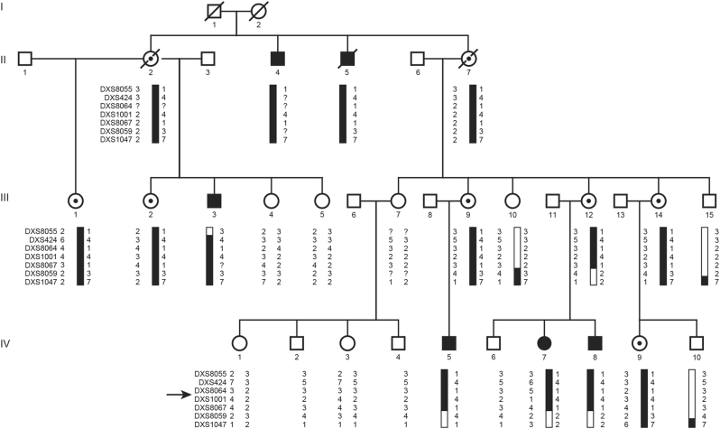
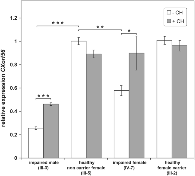
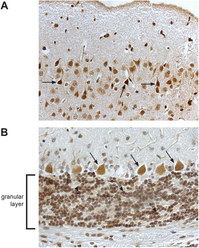
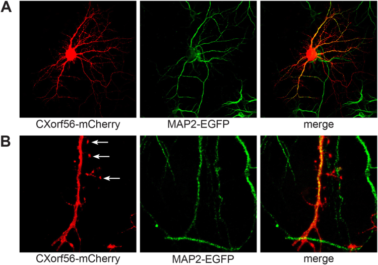
Intellectual disability (ID) comprises a large group of heterogeneous disorders, often without a known molecular cause. X-linked ID accounts for 5-10% of male ID cases. We investigated a large, three-generation family with mild ID and behavior problems in five males and one female, with a segregation suggestive for X-linked inheritance. Linkage analysis mapped a disease locus to a 7.6 Mb candidate region on the X-chromosome (LOD score 3.3). Whole-genome sequencing identified a 2 bp insertion in exon 2 of the chromosome X open reading frame 56 gene (CXorf56), resulting in a premature stop codon. This insertion was present in all intellectually impaired individuals and carrier females. Additionally, X-inactivation status showed skewed methylation patterns favoring the inactivation of the mutated allele in the unaffected carrier females. We demonstrate that the insertion leads to nonsense-mediated decay and that CXorf56 mRNA expression is reduced in the impaired males and female. In murine brain slices and primary hippocampal neuronal cultures, CXorf56 protein was present and localized in the nucleus, cell soma, dendrites, and dendritic spines. Although no other families have been identified with pathogenic variants in CXorf56, these results suggest that CXorf56 is the causative gene in this family, and thus a novel candidate gene for X-linked ID with behavior problems.


Conflict of interest statement

The authors declare that they have no conflict of interest.

Figures

Fig. 1
Fig. 1
The four-generation XLID pedigree and haplotype analysis results on Xq24. The disease-linked haplotype is depicted in black. Haplotypes are indicated with X-chromosome STR markers. The proximal boundary is determined by a recombination event in individual III-3 between DXS8055 and DXS424. The distal border is determined by both a recombination event in individual IV-5 and a historical recombination in IV-7 and IV-8 that was already present in III-12 between DXS8067 and DXS8059. An arrow indicates the location of the CXorf56 gene. Black symbols represent XLID impaired individuals, open symbols represent unaffected individuals, dotted symbols represent unaffected carriers, diagonal slash means deceased
Fig. 2
Fig. 2
Histogram displaying results of the relative quantitative CXorf56 mRNA analysis. Mean CXorf56 mRNA level relative to the reference gene GUSB and a calibrator sample (healthy non-carrier female (III-5)) are shown (±SEM). White and gray bars represent CXorf56 expression in EBV cell lines without (−CH) and with (+CH) treatment of cycloheximide, respectively. In the cell lines from an impaired male (III-3) and impaired female (IV-7) endogenous CXorf56 expression is significantly lower. After treatment with cycloheximide expression is significantly increased. Expression levels of an unaffected female carrier (III-2) are comparable to the control sample (III-5). *p < 0.05; **p < 0.01; ***p < 0.001
Fig. 3
Fig. 3
Subcellular localization of endogenous CXorf56 protein in murine brain. Immunohistochemical staining of endogenous CXorf56 protein in brain slices of 8-week-old male mice. Signal is present in the nucleus and cell soma of a pyramidal neurons in the cortex (examples indicated by black arrows) and b Purkinje cells (examples indicated by black arrows) and neurons in the granular layer (examples indicated by arrowheads) in the cerebellum. Note the nuclear as well as the cytoplasmic signal in the neurons
Fig. 4
Fig. 4
Localization of CXorf56 in cultured primary hippocampal neurons. ab Primary hippocampal neuron cultures of mice E17 pups. a Colocalization of CXorf56 protein in nucleus and dendrites (red, left panel) with endogenous Map2, a dendritic marker (green, middle panel), after 14 days in vitro. Merge (right panel). b High magnification of a dendrite with spines. Note the presence of CXorf56 protein in dendritic spines (indicated with arrows)

References

    1. Vissers LE, Gilissen C, Veltman JA. Genetic studies in intellectual disability and related disorders. Nat Rev Genet. 2016;17:9–18. doi: 10.1038/nrg3999. - DOI - PubMed
    1. Willemsen MH, Kleefstra T. Making headway with genetic diagnostics of intellectual disabilities. Clin Genet. 2014;85:101–10. doi: 10.1111/cge.12244. - DOI - PubMed
    1. Gecz J, Shoubridge C, Corbett M. The genetic landscape of intellectual disability arising from chromosome X. Trends Genet. 2009;25:308–16. doi: 10.1016/j.tig.2009.05.002. - DOI - PubMed
    1. Lubs HA, Stevenson RE, Schwartz CE. Fragile X and X-linked intellectual disability: four decades of discovery. Am J Hum Genet. 2012;90:579–90. doi: 10.1016/j.ajhg.2012.02.018. - DOI - PMC - PubMed
    1. Ropers HH. Genetics of early onset cognitive impairment. Annu Rev Genom Hum Genet. 2010;11:161–87. doi: 10.1146/annurev-genom-082509-141640. - DOI - PubMed