Review
. 2018 Jan 27;17(1):15.
doi: 10.1186/s12943-018-0772-6.

Extracellular vesicle-derived DNA for performing EGFR genotyping of NSCLC patients

Affiliations
Review

Extracellular vesicle-derived DNA for performing EGFR genotyping of NSCLC patients

Jae Young Hur et al. Mol Cancer. .

Abstract

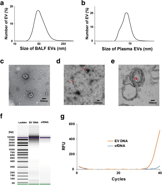
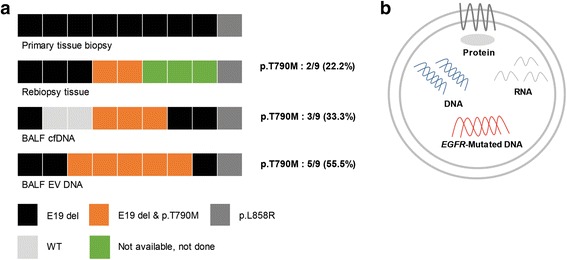
Tumor cells shed an abundance of extracellular vesicles (EVs) to body fluids containing bioactive molecules including DNA, RNA, and protein. Investigations in the field of tumor-derived EVs open a new horizon in understanding cancer biology and its potential as cancer biomarkers as well as platforms for personalized medicine. This study demonstrates that successfully isolated EVs from plasma and bronchoalveolar lavage fluid (BALF) of non-small cell lung cancer (NSCLC) patients contain DNA that can be used for EGFR genotyping through liquid biopsy. In both plasma and BALF samples, liquid biopsy results using EV DNA show higher accordance with conventional tissue biopsy compared to the liquid biopsy of cfDNA. Especially, liquid biopsy with BALF EV DNA is tissue-specific and extremely sensitive compared to using cfDNA. Furthermore, use of BALF EV DNA also demonstrates higher efficiency in comparison to tissue rebiopsy for detecting p.T790 M mutation in the patients who developed resistance to EGFR-TKIs. These finding demonstrate possibility of liquid biopsy using EV DNA potentially replacing the current diagnostic methods for more accurate, cheaper, and faster results.

Keywords: Bronchoalveolar lavage fluid; EGFR mutant DNA; Extracellular vesicles; Liquid biopsy; Non-small cell lung cancer.


Conflict of interest statement

Ethics approval and consent to participate

All human samples are collected under Institutional Review Committee (IRB).

Consent for publication

Not applicable.

Competing interests

The authors declare that they have no competing interests.

Publisher’s Note

Springer Nature remains neutral with regard to jurisdictional claims in published maps and institutional affiliations.

Figures

Fig. 1
Fig. 1
Characterization of BALF and plasma EV DNA. a. Size distribution of BALF EV. BALF was ultracentrifuged to obtain pallets and remove cells and debris, which was resuspended in 200 μl PBS. Sizes of purified EVs were determined using Zetasizer Nano ZS. Average size distribution from six separate experiments is plotted in percentage distribution according to their size. All six distributions are shown in Additional file 1: Figure S1. b. Size distribution of plasma EV. Plasma was ultracentrifuged to obtain pallets and remove cells and debris, which was resuspended in 200 μl PBS. Sizes of purified EVs were determined using Zetasizer Nano ZS. Average size distribution from three separate experiments is plotted in percentage distribution according to their size. All three distributions are shown in Additional file 1: Figure S2. c. EM image of BALF EVs. Samples for EM analysis were negatively stained. The size bar in the EM image indicates 100 nm. d. EM image of plssma EVs. Samples for EM analysis were negatively stained. Plasma EVs indicated by red arrows. The size bar in the EM image indicates 100 nm. e. Detection of dsDNA in BALF EVs by performing immuno-EM. DsDNA was labeled with a mouse monoclonal antibody and colloidal gold-conjugated secondary antibodies. The solid black dots indicate DNA (indicated by red arrows). f. Gel-like images show the size and amount of EV DNA and cfDNA determined using the bioanalyzer. First lane shows the standard size ladder distribution, and numbers on the left indicate corresponding sizes. The second and third lanes show the size and amount of EV DNA and cfDNA, respectively. g. Amplification curve obtained by performing real-time PCR. Exon 19 deletion in EGFR was determined by performing peptide nucleic acid (PNA)-mediated PCR clamping. Both EV DNA and cfDNA were extracted from 1 ml BALF, and 70 ng EV DNA and cfDNA were used for performing PCR
Fig. 2
Fig. 2
Detection of EGFR mutation by using BALF EV DNA. a. Comparison of EGFR genotyping results performed using tumor tissue, BALF cfDNA, and BALF EV DNA. The top lane indicates EGFR mutation status in the primary tumor tissue of each patient. The second lane shows the result of biopsy of tumor tissues obtained from patients with acquired resistance to EGFR-TKIs. The third and fourth lanes show EGFR mutation status determined by performing liquid biopsy of BALF cfDNA and EV DNA, respectively. Rates of p.T790 M mutation detection by performing tissue rebiopsy and liquid biopsy of BALF cfDNA and BALF EV DNA are shown on the right side of each lane. Abbreviations; E19 del: exon 19 deletion, WT: wild type. b. Diagram showing the contents of an EV. Tumor-derived EVs contain RNA; DNA, including mutant DNA; and proteins

Similar articles

Cited by

References

    1. Reck M, Rabe KF. Precision diagnosis and treatment for advanced non-small-cell lung cancer. N Engl J Med. 2017;377:849–861. doi: 10.1056/NEJMra1703413. - DOI - PubMed
    1. Huang WL, Chen YL, Yang SC, Ho CL, Wei F, Wong DT, Su WC, Lin CC. Liquid biopsy genotyping in lung cancer: ready for clinical utility? Oncotarget. 2017;8:18590–18608. - PMC - PubMed
    1. Molina-Vila MA. Liquid biopsy in lung cancer: present and future. Transl Lung Cancer Res. 2016;5:452–454. doi: 10.21037/tlcr.2016.10.05. - DOI - PMC - PubMed
    1. Kohler C, Radpour R, Barekati Z, Asadollahi R, Bitzer J, Wight E, Burki N, Diesch C, Holzgreve W, Zhong XY. Levels of plasma circulating cell free nuclear and mitochondrial DNA as potential biomarkers for breast tumors. Mol Cancer. 2009;8:105. doi: 10.1186/1476-4598-8-105. - DOI - PMC - PubMed
    1. Kahlert C, Melo SA, Protopopov A, Tang J, Seth S, Koch M, Zhang J, Weitz J, Chin L, Futreal A, Kalluri R. Identification of double-stranded genomic DNA spanning all chromosomes with mutated KRAS and p53 DNA in the serum exosomes of patients with pancreatic cancer. J Biol Chem. 2014;289:3869–3875. doi: 10.1074/jbc.C113.532267. - DOI - PMC - PubMed

Publication types