. 2018 Jan 11:8:970.
doi: 10.3389/fphar.2017.00970. eCollection 2017.

Cisplatin and Pemetrexed Activate AXL and AXL Inhibitor BGB324 Enhances Mesothelioma Cell Death from Chemotherapy

Affiliations

Cisplatin and Pemetrexed Activate AXL and AXL Inhibitor BGB324 Enhances Mesothelioma Cell Death from Chemotherapy

Derek B Oien et al. Front Pharmacol. .

Abstract

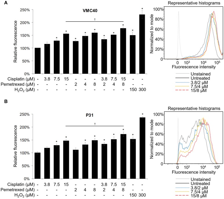
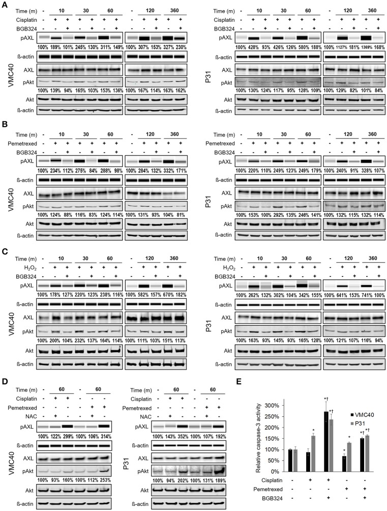
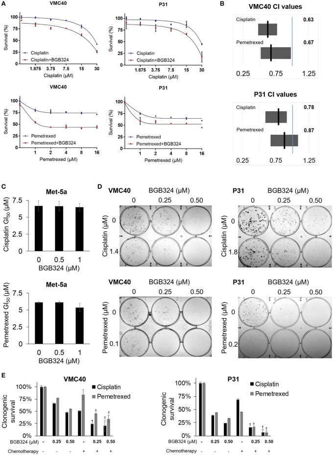
Reactive oxygen species (ROS) can promote or inhibit tumorigenesis. In mesothelioma, asbestos exposure to serous membranes induces ROS through iron content and chronic inflammation, and ROS promote cell survival signaling in mesothelioma. Moreover, a current chemotherapy regimen for mesothelioma consisting of a platinum and antifolate agent combination also induce ROS. Mesothelioma is notoriously chemotherapy-resistant, and we propose that ROS induced by cisplatin and pemetrexed may promote cell survival signaling pathways, which ultimately may contribute to chemotherapy resistance. In The Cancer Genome Atlas datasets, we found AXL kinase expression is relatively high in mesothelioma compared to other cancer samples. We showed that ROS induce the phosphorylation of AXL, which was blocked by the selective inhibitor BGB324 in VMC40 and P31 mesothelioma cells. We also showed that cisplatin and pemetrexed induce the phosphorylation of AXL and Akt, which was also blocked by BGB324 as well as by N-acetylcysteine antioxidant. AXL knockdown in these cells enhances sensitivity to cisplatin and pemetrexed. Similarly, AXL inhibitor BGB324 also enhances sensitivity to cisplatin and pemetrexed. Finally, higher synergy was observed when cells were pretreated with BGB324 before adding chemotherapy. These results demonstrate cisplatin and pemetrexed induce ROS that activate AXL, and blocking AXL activation enhances the efficacy of cisplatin and pemetrexed. These results suggest AXL inhibition combined with the current chemotherapy regimen may represent an effective strategy to enhance the efficacy of chemotherapy in mesothelioma. This is the first study, to our knowledge, on chemotherapy-induced activation of AXL and cell survival pathways associated with ROS signaling.

Keywords: AXL; BGB324; chemotherapy resistance; cisplatin; mesothelioma; pemetrexed; reactive oxygen species.


Figures

Figure 1
Figure 1
AXL is abnormally expressed in MPM. (A) AXL and GAS6 expression are elevated in clinical MPM samples. Of 30 TCGA cancer datasets, MPM (87 samples, blue dots) ranked fourth highest for AXL and third highest for GAS6 expression, and mean log2 values were 11.40 and 13.06, respectively. Data are RNA Seq V2 values from TCGA Research Network. (B) Reverse phase protein heat map subset with AXL. MPM cell extracts applied to coated slides and probed with 301 antibodies, normalized to loading. Color brightness indicates protein detection relative to median, red for increased and green for decreased detection levels. Normalized log2 median-centered value of AXL was 1.35 for VMC40. (C) Lysates analyzed for pAXL and GAS6 ligand using capillary electrophoresis, and other proteins were detected by standard immunoblot methods. Met-5a as a nonmalignant mesothelial control cell line for comparison. Densitometry values are relative to corresponding protein of Met-5a, normalized to respective ß-actin loading control.
Figure 2
Figure 2
Cisplatin and pemetrexed induce ROS generation. The CM-H2DCFDA ROS indicator was preloaded into (A) VMC40 and (B) P31 cell lines, followed by 30 m of cisplatin and pemetrexed treatment. Fluorescent signal was detected by flow cytometry, and area-under-curve values were compared to untreated samples as percentages. Representative histograms of combined cisplatin and pemetrexed concentrations on right. Error bars represent ±SEM of three experiments. * and † denote p < 0.05 via t-test for value compared to untreated control and to drug combination, respectively.
Figure 3
Figure 3
Cisplatin and pemetrexed promote phosphorylation of AXL and downstream Akt. (A–C) Time course of pAXL and downstream pAkt detection in the presence of (A) 15 μM cisplatin, (B) 8 μM pemetrexed, and (C) 150 μM H2O2. Phosphorylation of AXL was inhibited by adding 2 μM BGB324. Densitometry values as percentage of untreated control are shown for pAXL and pAkt, normalized to respective beta-actin. (D) pAXL detection with the addition of 4 mM N-acetylcysteine (NAC) antioxidant at 60 m. Lysates (A–D) were analyzed for pAXL using capillary electrophoresis, and other proteins were detected by standard immunoblot methods. Densitometry values are relative to corresponding protein of untreated control, normalized to respective ß-actin loading control. (E) Caspase 3 activity after 24 h of indicated drug treatment. Error bars represent ±SEM of three experiments. * and † denote p < 0.05 via t-test for value compared to untreated control and to drug combination, respectively.
Figure 4
Figure 4
AXL knockdown enhances cisplatin and pemetrexed efficacy. (A) Relative quantification of AXL after shRNA AXL knockdown using three gel loading volumes of 5, 15, and 30 μL. Values are percentage of NTC. (B) Cell proliferation via SRB assay, values compared as percentage to NTC after 4 days of plating equal cell numbers. *Denotes p < 0.05 via t-test for value compared to NTC value. (C) Cell survival percentages by SRB assay after 72 h treatment with cisplatin (top) and pemetrexed (bottom) at incrementing concentrations. (D) Cell survival percentages by clonogenic assay after 72 h treatment with cisplatin (top) and pemetrexed (bottom) at incrementing concentrations. Values are relative to no drug treatment (100% for both NTC and AXL shRNA). †Denotes p < 0.05 via t-test for value compared to NTC value at the same concentration. Error bars represent ±SEM of three experiments (B-D).
Figure 5
Figure 5
Combination of BGB324 with cisplatin and pemetrexed enhances MPM cell death. (A) Cell survival for incrementing concentrations of cisplatin and pemetrexed with and without 0.5 μM BGB324. Values determined by SRB assay at 72 h. Actual values, dose-response curve of BGB324, and statistical analysis at specified concentrations are shown in Supplementary Figure 7. (B) CI range (gray box) and mean (black line) from full data shown in Supplementary Figure 7A. The reference value, 1.00, is indicated by a blue line. CI < 1.00 is considered synergistic. CI values calculated as described in section Materials and Methods. (C) GI50 values for cisplatin and pemetrexed in nonmalignant Met-5a cells. (D) Representative images of clonogenic assays after 72 h treatment and then allowing cells to form colonies in absence of drugs. (E) Quantification of clonogenic assays. †Denotes p < 0.05 via t-test for value compared to values of single agents. Error bars represent ±SEM of three experiments (A,C,E).
Figure 6
Figure 6
Time-sequenced BGB324 combination with cisplatin and pemetrexed enhances MPM cell death. BGB324 was added 4 h before other drugs. (A) Cell survival for incrementing concentrations of cisplatin and pemetrexed with and without 0.5 μM BGB324. Values determined by SRB assay at 72 h. Actual values, incrementing concentrations of BGB324, and statistical analysis at specified concentrations are shown in Supplementary Figure 8. (B) CI range (gray box) and mean (black line) from full data shown in Supplementary Figure 8A. The reference value, 1.00, is indicated by a blue line. CI < 1.00 is considered synergistic. CI values calculated as described in section Materials and Methods. (C) GI50 values for cisplatin and pemetrexed in nonmalignant Met-5a cells. (D) Representative images of clonogenic assays after 72 h treatment and then allowing cells to form colonies in absence of drugs. (E) Quantification of clonogenic assays. †Denotes p < 0.05 via t-test for value compared to values of single agents. Error bars represent ±SEM of three experiments (A,C,E).
Figure 7
Figure 7
Combination of BGB324 with both cisplatin and pemetrexed. (A) Representative images of clonogenic assays after 72 h treatment with BGB324, cisplatin, and pemetrexed. (B) Representative images of clonogenic assays after 4-h pretreatment with BGB324 followed 72 h treatment with both cisplatin and pemetrexed. (C) Quantification of (A,B) by measuring quantity of stain from cells. †Denotes p < 0.05 via t-test for value compared to values of BGB324 alone. Error bars represent ±SEM of three experiments.
Figure 8
Figure 8
Known pathways and hypothesis for AXL inhibitor (red box) and platinum or antifolate drug (blue box) synergies. Black arrows: Activated AXL promotes cell survival. ROS can increase GAS6 activation and activate AXL independently. Cisplatin and pemetrexed cause cell cycle arrest and increased ROS promoting apoptosis, but ROS-induced AXL activation can inhibit apoptosis. Red line: AXL inhibition can decrease cell survival and promote apoptosis. We have shown adding an AXL inhibitor to current chemotherapy will synergistically promote cell death. This synergy is based on inhibiting an opposing pathway of apoptosis.

Similar articles

Cited by

References

    1. Akaboshi M., Kawai K., Maki H., Akuta K., Ujeno Y., Miyahara T. (1992). The number of platinum atoms binding to DNA, RNA and protein molecules of HeLa cells treated with cisplatin at its mean lethal concentration. Jpn. J. Cancer Res. 83, 522–526. 10.1111/j.1349-7006.1992.tb01959.x - DOI - PMC - PubMed
    1. Andersson A., Fagerberg J., Lewensohn R., Ehrsson H. (1996). Pharmacokinetics of cisplatin and its monohydrated complex in humans. J. Pharm. Sci. 85, 824–827. 10.1021/js960037a - DOI - PubMed
    1. Azar Z. M., Mehdi M. Z., Srivastava A. K. (2007). Insulin-like growth factor type-1 receptor transactivation in vasoactive peptide and oxidant-induced signaling pathways in vascular smooth muscle cells. Can. J. Physiol. Pharmacol. 85, 105–11. 10.1139/Y06-101 - DOI - PubMed
    1. Bae S. Y., Hong J. Y., Lee H. J., Park H. J., Lee S. K. (2015). Targeting the degradation of AXL receptor tyrosine kinase to overcome resistance in gefitinib-resistant non-small cell lung cancer. Oncotarget 6, 10146–10160. 10.18632/oncotarget.3380 - DOI - PMC - PubMed
    1. Baregamian N., Song J., Chung D. H. (2012). Effects of oxidative stress on intestinal type I insulin-like growth factor receptor expression. Eur. J. Pediatr. Surg. 22, 97–104. 10.1055/s-0032-1306261 - DOI - PMC - PubMed