. 2018 Jan 11:8:2540.
doi: 10.3389/fmicb.2017.02540. eCollection 2017.

Virome Characterization of a Collection of S. sclerotiorum from Australia

Affiliations

Virome Characterization of a Collection of S. sclerotiorum from Australia

Fan Mu et al. Front Microbiol. .

Abstract

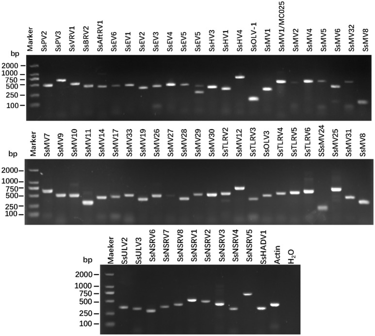
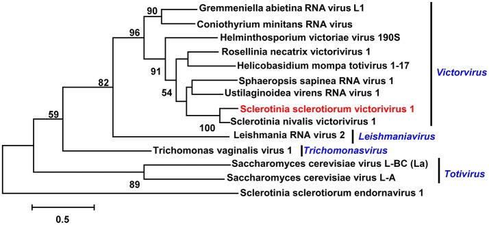
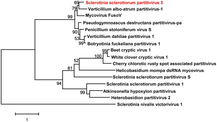
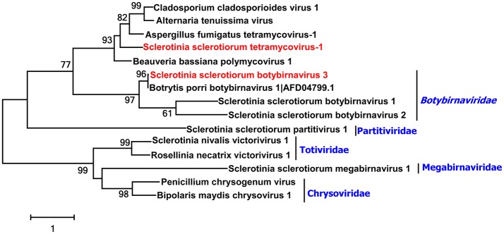
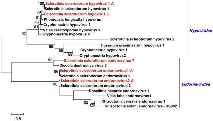
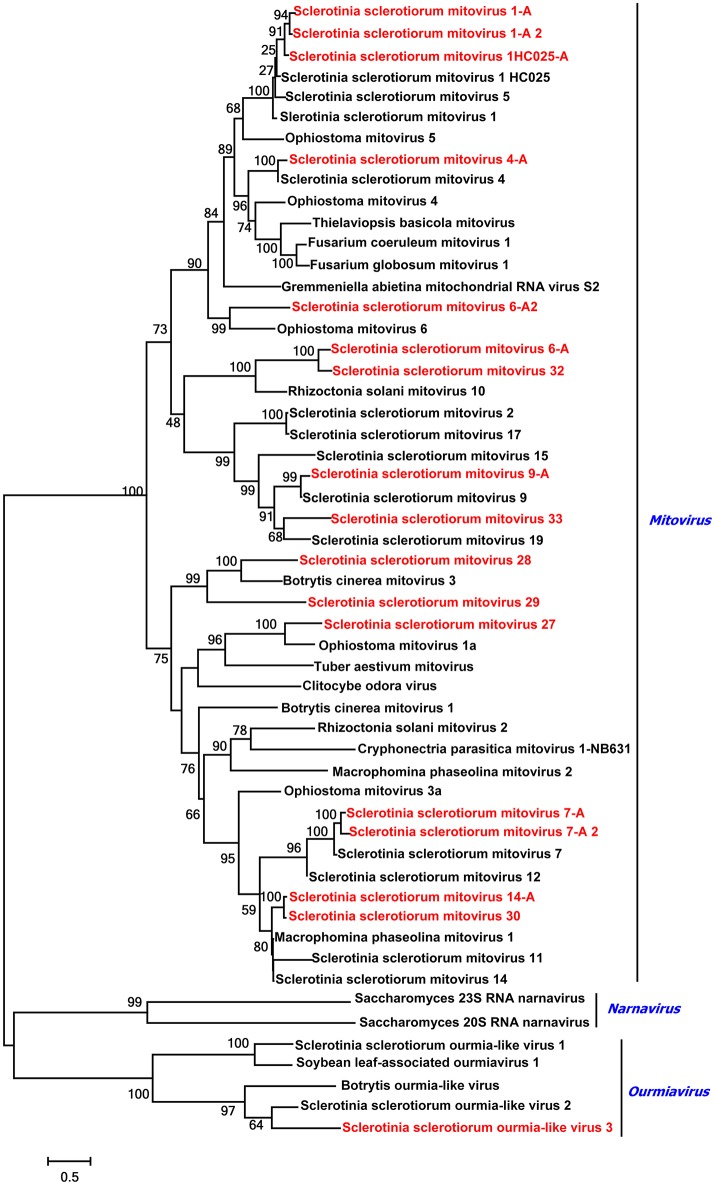
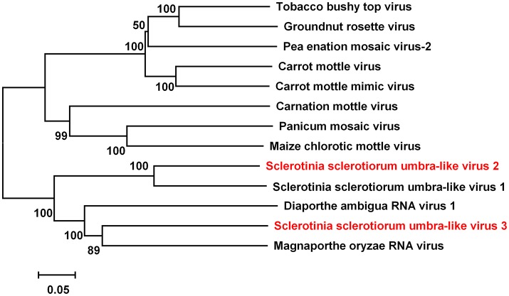
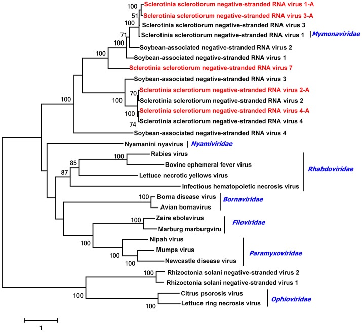
Sclerotinia sclerotiorum is a devastating plant pathogen that attacks numerous economically important broad acre and vegetable crops worldwide. Mycoviruses are widespread viruses that infect fungi, including S. sclerotiorum. As there were no previous reports of the presence of mycoviruses in this pathogen in Australia, studies were undertaken using RNA_Seq analysis to determine the diversity of mycoviruses in 84 Australian S. sclerotiorum isolates collected from various hosts. After RNA sequences were subjected to BLASTp analysis using NCBI database, 285 contigs representing partial or complete genomes of 57 mycoviruses were obtained, and 34 of these (59.6%) were novel viruses. These 57 viruses were grouped into 10 distinct lineages, namely Endornaviridae (four novel mycoviruses), Genomoviridae (isolate of SsHADV-1), Hypoviridae (two novel mycoviruses), Mononegavirales (four novel mycovirusess), Narnaviridae (10 novel mycoviruses), Partitiviridae (two novel mycoviruses), Ourmiavirus (two novel mycovirus), Tombusviridae (two novel mycoviruses), Totiviridae (one novel mycovirus), Tymovirales (five novel mycoviruses), and two non-classified mycoviruses lineages (one Botrytis porri RNA virus 1, one distantly related to Aspergillus fumigatus tetramycovirus-1). Twenty-five mitoviruses were determined and mitoviruses were dominant in the isolates tested. This is not only the first study to show existence of mycoviruses in S. sclerotiorum in Australia, but highlights how they are widespread and that many novel mycoviruses occur there. Further characterization of these mycoviruses is warranted, both in terms of exploring these novel mycoviruses for innovative biocontrol of Sclerotinia diseases and in enhancing our overall knowledge on viral diversity, taxonomy, ecology, and evolution.

Keywords: Australia; RNA_sequencing; Sclerotinia sclerotiorum; biocontrol; mycovirus; virome; virus diversity.


Figures

Figure 1
Figure 1
RT-PCR confirmation of mycovirus contigs. RT-PCR confirmation of de novo assembled mycovirus contigs from Sclerotinia sclerotiorum generated by Illumina sequencing. The presences of viral sequences identified by BLAST analysis were confirmed by RT-PCR products generated from total RNA samples. The primers were designed according to the contigs' sequences (genomic sequences of putative mycoviruses). Primers pairs used and predicted sizes of amplicons are listed in Table S3. Lane M, DNA marker, 2,000 bp DNA Ladder (Takara Bio Inc., Japan); Lane 1 to 58, abbreviates of viruses (see Table 1 for detail), Lane H2O, ddH2O was instead of RT products as control.
Figure 2
Figure 2
Phylogenetic analysis of the putative virus in Family Totiviridae. Phylogenetic analysis of the putative Sclerotinia sclerotiorum victorivirus 1 with other selected viruses in Family Totiviridae based on viral RdRp amino acid sequences. Branch lengths are scaled to the expected underlying number of amino acid substitutions per site. If not specifically addressed, the alignments of RdRp amino acid sequences were performed by ClustalW, and phylogenetic tree was constructed using MEGA7.0.18, and selected viruses are listed in Table S4. Viruses marked with red color are found in Australian isolates of Sclerotinia sclerotiorum. Sclerotinia sclerotiorum endornavirus 1 was as the outgroup.
Figure 3
Figure 3
Phylogenetic analysis of Sclerotinia sclerotiorum partitivirus. Phylogenetic analysis of Sclerotinia sclerotiorum partitivirus 3 and other selected viruses based on the RdRp amino acid sequences. Viruses marked with red color are found in Australian isolates of Sclerotinia sclerotiorum. Sclerotinia nivalis victorivirus 1 was as the outgroup.
Figure 4
Figure 4
Phylogenetic analysis of two unclassified viruses. Phylogenetic analysis of Sclerotinia sclerotiorum tetramycovirus-1 and Sclerotinia sclerotiorum botybirnavirus 3 with other selected viruses based on the RdRp protein amino acid sequences. Viruses marked with red color are found in Australian isolates of Sclerotinia sclerotiorum.
Figure 5
Figure 5
Phylogenetic analysis of viruses in the Family Endornaviridae and Hypoviridae. Phylogenetic analysis of viruses in the Family Endornaviridae and viruses in the Family Hypoviridae based on multiple alignments of full-length RdRp amino acid sequences. Viruses marked with red color are found in Australian isolates of Scleorotinia sclerotiorum.
Figure 6
Figure 6
Phylogenetic analysis of Mitoviruses and Ourmiavirus. Phylogenetic relationships of mitoviruses and ourmiavirus that infect Sclerotinia sclerotiorum and other selected viruses. Multi alignments are based on the virus's full-length RdRp amino acid sequences. Viruses marked with red color are found in Australian isolates of Sclerotinia sclerotiorum.
Figure 7
Figure 7
Phylogenetic analysis of virus in the Family Tombusviridae. Phylogenetic relationships of putative new virus genomes of Tombusviridae virus. Viruses marked with red color are found in Australian isolates of Sclerotinia sclerotiorum.
Figure 8
Figure 8
Phylogenetic relationships of putative negative-stranded RNA virus genomes. For the neighbor joining tree, predicted RdRp amino acid sequences were aligned, and phylogenetic trees were constructed. Viruses marked with red color are found in Australian isolates of Sclerotinia sclerotiorum.

References

    1. Afonso C. L., Amarasinghe G. K., Bányai K., Bào Y., Basler C. F., Bavari S., et al. . (2016). Taxonomy of the order Mononegavirales: update 2016. Arch. Virol. 161, 2351–2360. 10.1007/s00705-016-2880-1 - DOI - PMC - PubMed
    1. Barbetti M. J., Banga S. K., Fu T. D., Li Y. C., Singh D., Liu S. Y., et al. (2014). Comparative genotype reactions to Sclerotinia sclerotiorum within breeding populations of Brassica napus and B. juncea from India and China. Euphytica 197, 47–59. 10.1007/s10681-013-1049-1 - DOI
    1. Bolton M. D., Thomma B. P., Nelson B. D. (2006). Sclerotinia sclerotiorum (Lib.) de Bary: biology and molecular traits of a cosmopolitan pathogen. Mol. Plant Pathol. 7:1. 10.1111/j.1364-3703.2005.00316.x - DOI - PubMed
    1. Bretag T. W., Mebalds M. I., Bretag T. W., Mebalds M. I. (1987). Pathogenicity of fungi isolated from Cicer arietinum (chickpea) grown in north-western Victoria. Aust. J. Exp. Agric. 27, 141–148. 10.1071/EA9870141 - DOI
    1. Bsm L., Ochoacorona F. M., Elliott D. R., Tang J. Z., Bjr A. (2007). Detection of Poinsettia mosaic virus by RT-PCR in Euphorbia spp. in New Zealand. Plant Dis. 91, 110–110. 10.1094/PD-91-0110A - DOI - PubMed