Review
. 2018 Jan 9:8:2634.
doi: 10.3389/fmicb.2017.02634. eCollection 2017.

Probing the Structures of Viral RNA Regulatory Elements with SHAPE and Related Methodologies

Affiliations
Review

Probing the Structures of Viral RNA Regulatory Elements with SHAPE and Related Methodologies

Jason W Rausch et al. Front Microbiol. .

Abstract

Viral RNAs were selected by evolution to possess maximum functionality in a minimal sequence. Depending on the classification of the virus and the type of RNA in question, viral RNAs must alternately be replicated, spliced, transcribed, transported from the nucleus into the cytoplasm, translated and/or packaged into nascent virions, and in most cases, provide the sequence and structural determinants to facilitate these processes. One consequence of this compact multifunctionality is that viral RNA structures can be exquisitely complex, often involving intermolecular interactions with RNA or protein, intramolecular interactions between sequence segments separated by several thousands of nucleotides, or specialized motifs such as pseudoknots or kissing loops. The fluidity of viral RNA structure can also present a challenge when attempting to characterize it, as genomic RNAs especially are likely to sample numerous conformations at various stages of the virus life cycle. Here we review advances in chemoenzymatic structure probing that have made it possible to address such challenges with respect to cis-acting elements, full-length viral genomes and long non-coding RNAs that play a major role in regulating viral gene expression.

Keywords: RNA structure; SHAPE; chemical probing; secondary structure; viral RNA.


Figures
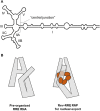
Figure 1
Figure 1
(A) Cartoon representation of a 5-SL form of the HIV-1 RRE with basepairing in the central junction. Stem, stem loop and central junction designations are indicated. (B) SAXS, SHAPE and aiSHAPE analysis suggests that the distal segment of stem I (S-I) folds back on itself to provide a cryptic Rev binding site, pre-organizing the RRE for optimal Rev multimerization. Three-dimensional RRE and Rev6-RRE complex model structures are shown. Adapted from Bai et al. (2014).
Figure 2
Figure 2
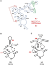
(A) Secondary structure of the musD, MTE, indicating long range interactions important for nucleocytoplasmic RNA transport. An annotated secondary structure is provided, where S, L, SL, and IL refer to stem, loop, stem-loop and internal loop, respectively. The L3/IL8 kissing interaction is indicated in red and the S13/SL12 pseudoknot in green. (B) Site-directed mutagenesis that interrupts the former interaction results in loss of MTE function (mutant M4). (C) In contrast, creating a substitute kissing loop interaction (mutant M5), restores activity, suggesting the importance of the kissing interaction in supporting overall MTE topology. See Legiewicz et al. (2010) for additional details on MTE function and mutagenesis.
Figure 3
Figure 3
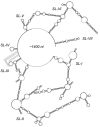
SHAPE-predicted secondary structure analysis of the ~1,400 nt gammaretroviral RNA transport element, PTE. Stem-loops (SL) SL-I through SL-VII are indicated. Proposed long range interactions involving nucleotides of SL-III are indicated by dotted lines. Adapted from Pilkington et al. (2014).
Figure 4
Figure 4
Resolving alternate conformers of the HIV-1 5′UTR by in-gel SHAPE. (A) Schematic depicting native polyacrylamide gel electrophoresis of in vitro-transcribed HIV-1 5′ UTR RNA to resolve the monomer (M) and dimer species (D), which are excised and treated with the SHAPE reagent in situ. (B) Following recovery of chemically modified RNAs, the SHAPE protocol is completed, revealing the monomeric and dimeric configurations of the 5'UTR construct. Adapted from Kenyon et al. (2013).
Figure 5
Figure 5
Dissecting alternative topologies for the HIV-1 RRE by native gel electrophoresis and in-gel SHAPE. The central panel illustrates that the RRE exists as an equilibrium mixture of two conformers that can be separated by prolonged electrophoresis. In-gel SHAPE indicates that the faster and slower migrating forms assume the 4-SL and 5-SL conformations, respectively. Adapted from Sherpa et al. (2015).
Figure 6
Figure 6
Analysis of HIV-2 RRE conformational changes during folding. In vitro transcribed RNA constructs are thermally denatured, flash cooled to 4°C and then incubated at 37°C. Immediately after flash cooling, the HIV-2 RRE exists as a mixture of A, B, and C conformations. Upon raising the temperature to 37°C, the relative ratios of the three variants changes over time, with conformer C ultimately predominating. (A) Schematic depicting non-denaturing gel electrophoresis of HIV-2 RRE as a function of incubation time. (B) Fluorometric quantification of fractionated RRE conformers. (C) Proposed models of the HIV-2 RRE open (Conformer A), intermediate (Conformer B) and closed forms (Conformer C). Secondary structural motifs are indicated and color-coded as follows: SL I, red; SL IIA, dark green, SL IIB, IIC and adjacent connecting loops, magenta; SL III, yellow; SL IV, blue; SL V, orange. Adapted from Lusvarghi et al. (2013a).
Figure 7
Figure 7
Secondary structure of the DENV minigenome determined by CE-SHAPE. Stem loop A (SLA), dumbbell, pseudoknot (PK1 and PK2), terminal loop (TL1), capsid-coding region hairpin element (cHP) and 5′-3′ interaction motifs are indicated. Despite the designation, neither CE-SHAPE nor aiSHAPE indicated formation of a pseudoknot involving the PK1 motif. Nucleotide shading is in proportion to normalized reactivity. Adapted from Sztuba-Solinska et al. (2013).
Figure 8
Figure 8
Secondary structure of the EBOV minigenome determined by CE-SHAPE. Predicted base pairing of 3′ leader and 5′ trailer sequences is shown together with a heat shock protein A 8 (HSPA 8) binding site. The discontinuity created by omission of the GFP gene from the displayed minigenome construct sequence is also indicated. Nucleotide shading is in proportion to normalized reactivity. Adapted from Sztuba-Solinska et al. (2016).
Figure 9
Figure 9
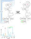
A kissing loop motif within the musD RNA transport element validated by aiSHAPE. (A) Predicted secondary structure the musD RNA transport element, including a postulated kissing loop interaction between nt 82–89 and nt 182–189. (B) The LNA-DNA chimera designed to hybridize to nt 182–189 would be expected to displace nt 82–89 and render them more susceptible to acylation. Perturbation of RNA structure outside of this region should be minimal. (C) A step plot comparing RNA reactivity values obtained in the presence and absence of the LNA-DNA chimera. A marked increase in reactivity was observed for nt 82–89 in the presence of the chimera, thus validating the kissing loop prediction. Adapted from Legiewicz et al. (2010).
Figure 10
Figure 10
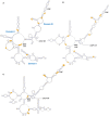
PAN RNA secondary structures and potential KSHV protein binding sites. Models generated from SHAPE-MaP experiments using PAN RNA isolated from (A) nuclei, (B) cytoplasm or (C) virions. The MRE core motif and poly-A tail are indicated in all panels, while Domains I-III are common to all three panels but are indicated only in panel (A). Orange zones reflect differences in reactivity calculated for RNA probed in- or ex-vivo that are consistent with protein binding. Adapted from Sztuba-Solinska et al. (2017).

Similar articles

Cited by

References

    1. Askjaer P., Jensen T. H., Nilsson J., Englmeier L., Kjems J. (1998). The specificity of the CRM1-Rev nuclear export signal interaction is mediated by RanGTP. J. Biol. Chem. 273, 33414–33422. 10.1074/jbc.273.50.33414 - DOI - PubMed
    1. Bai Y., Tambe A., Zhou K., Doudna J. A. (2014). RNA-guided assembly of Rev-RRE nuclear export complexes. Elife 3:e03656. 10.7554/eLife.03656 - DOI - PMC - PubMed
    1. Bechtel J., Grundhoff A., Ganem D. (2005). RNAs in the virion of Kaposi's sarcoma-associated herpesvirus. J. Virol. 79, 10138–10146. 10.1128/JVI.79.16.10138-10146.2005 - DOI - PMC - PubMed
    1. Bolton E. C., Coombes C., Eby Y., Cardell M., Boeke J. D. (2005). Identification and characterization of critical cis-acting sequences within the yeast Ty1 retrotransposon. RNA 11, 308–322. 10.1261/rna.7860605 - DOI - PMC - PubMed
    1. Booth D. S., Cheng Y., Frankel A. D. (2014). The export receptor Crm1 forms a dimer to promote nuclear export of HIV RNA. Elife 3:e04121. 10.7554/eLife.04121 - DOI - PMC - PubMed

LinkOut - more resources