

Nanoporous Microneedle Arrays Effectively Induce Antibody Responses against Diphtheria and Tetanus Toxoid
- PMID: 29375544
- PMCID: PMC5770646
- DOI: 10.3389/fimmu.2017.01789
Nanoporous Microneedle Arrays Effectively Induce Antibody Responses against Diphtheria and Tetanus Toxoid
Abstract
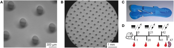
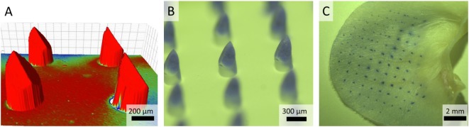
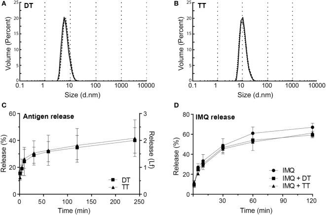
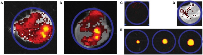
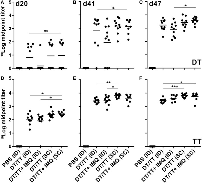
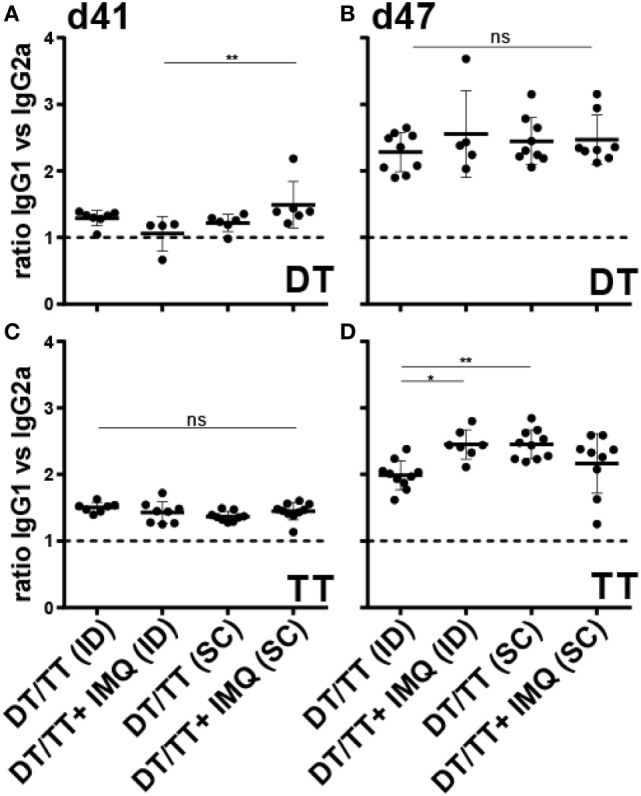
The skin is immunologically very potent because of the high number of antigen-presenting cells in the dermis and epidermis, and is therefore considered to be very suitable for vaccination. However, the skin's physical barrier, the stratum corneum, prevents foreign substances, including vaccines, from entering the skin. Microneedles, which are needle-like structures with dimensions in the micrometer range, form a relatively new approach to circumvent the stratum corneum, allowing for minimally invasive and pain-free vaccination. In this study, we tested ceramic nanoporous microneedle arrays (npMNAs), representing a novel microneedle-based drug delivery technology, for their ability to deliver the subunit vaccines diphtheria toxoid (DT) and tetanus toxoid (TT) intradermally. First, the piercing ability of the ceramic (alumina) npMNAs, which contained over 100 microneedles per array, a length of 475 µm, and an average pore size of 80 nm, was evaluated in mouse skin. Then, the hydrodynamic diameters of DT and TT and the loading of DT, TT, and imiquimod into, and subsequent release from the npMNAs were assessed in vitro. It was shown that DT and TT were successfully loaded into the tips of the ceramic nanoporous microneedles, and by using near-infrared fluorescently labeled antigens, we found that DT and TT were released following piercing of the antigen-loaded npMNAs into ex vivo murine skin. Finally, the application of DT- and TT-loaded npMNAs onto mouse skin in vivo led to the induction of antigen-specific antibodies, with titers similar to those obtained upon subcutaneous immunization with a similar dose. In conclusion, we show for the first time, the potential of npMNAs for intradermal (ID) immunization with subunit vaccines, which opens possibilities for future ID vaccination designs.
Keywords: antigen release; diphtheria; humoral immune response; intradermal vaccination; nanoporous microneedles; tetanus.
Figures
Similar articles
-
Vaccination with influenza hemagglutinin-loaded ceramic nanoporous microneedle arrays induces protective immune responses.Eur J Pharm Biopharm. 2019 Mar;136:259-266. doi: 10.1016/j.ejpb.2019.02.002. Epub 2019 Feb 5. Eur J Pharm Biopharm. 2019. PMID: 30731115
-
Diphtheria toxoid and N-trimethyl chitosan layer-by-layer coated pH-sensitive microneedles induce potent immune responses upon dermal vaccination in mice.J Control Release. 2017 Sep 28;262:28-36. doi: 10.1016/j.jconrel.2017.07.017. Epub 2017 Jul 11. J Control Release. 2017. PMID: 28710002
-
Coated and Hollow Microneedle-Mediated Intradermal Immunization in Mice with Diphtheria Toxoid Loaded Mesoporous Silica Nanoparticles.Pharm Res. 2018 Aug 13;35(10):189. doi: 10.1007/s11095-018-2476-4. Pharm Res. 2018. PMID: 30105542 Free PMC article.
-
Microneedles and other physical methods for overcoming the stratum corneum barrier for cutaneous gene therapy.Crit Rev Ther Drug Carrier Syst. 2006;23(3):205-58. doi: 10.1615/critrevtherdrugcarriersyst.v23.i3.20. Crit Rev Ther Drug Carrier Syst. 2006. PMID: 17206925 Review.
-
[Microneedle-based percutaneous immunity: a review].Sheng Wu Gong Cheng Xue Bao. 2022 Sep 25;38(9):3301-3315. doi: 10.13345/j.cjb.220142. Sheng Wu Gong Cheng Xue Bao. 2022. PMID: 36151801 Review. Chinese.
Cited by
-
Microneedles: An Emerging Vaccine Delivery Tool and a Prospective Solution to the Challenges of SARS-CoV-2 Mass Vaccination.Pharmaceutics. 2023 Apr 27;15(5):1349. doi: 10.3390/pharmaceutics15051349. Pharmaceutics. 2023. PMID: 37242591 Free PMC article. Review.
-
Microneedle Arrays Combined with Nanomedicine Approaches for Transdermal Delivery of Therapeutics.J Clin Med. 2021 Jan 6;10(2):181. doi: 10.3390/jcm10020181. J Clin Med. 2021. PMID: 33419118 Free PMC article. Review.
-
Microarray patches enable the development of skin-targeted vaccines against COVID-19.Adv Drug Deliv Rev. 2021 Apr;171:164-186. doi: 10.1016/j.addr.2021.01.022. Epub 2021 Feb 2. Adv Drug Deliv Rev. 2021. PMID: 33539853 Free PMC article. Review.
-
Evaluation of efficacy and safety of intradermal delivery of vaccines through microneedle(s) in human beings: a protocol for a systematic review.Syst Rev. 2022 Aug 13;11(1):170. doi: 10.1186/s13643-022-02046-8. Syst Rev. 2022. PMID: 35964062 Free PMC article.
-
The potential role of using vaccine patches to induce immunity: platform and pathways to innovation and commercialization.Expert Rev Vaccines. 2020 Feb;19(2):175-194. doi: 10.1080/14760584.2020.1732215. Epub 2020 Mar 17. Expert Rev Vaccines. 2020. PMID: 32182145 Free PMC article. Review.
References
-
- Zaric M, Lyubomska O, Poux C, Hanna ML, McCrudden MT, Malissen B, et al. Dissolving microneedle delivery of nanoparticle-encapsulated antigen elicits efficient cross-priming and th1 immune responses by murine langerhans cells. J Invest Dermatol (2015) 135(2):425–34.10.1038/jid.2014.415 - DOI - PubMed
LinkOut - more resources
Full Text Sources
Other Literature Sources