. 2017 Dec 22:8:1890.
doi: 10.3389/fimmu.2017.01890. eCollection 2017.

Antibiotic-Induced Pathobiont Dissemination Accelerates Mortality in Severe Experimental Pancreatitis

Affiliations

Antibiotic-Induced Pathobiont Dissemination Accelerates Mortality in Severe Experimental Pancreatitis

Fernanda S Soares et al. Front Immunol. .

Abstract

Although antibiotic-induced dysbiosis has been demonstrated to exacerbate intestinal inflammation, it has been suggested that antibiotic prophylaxis may be beneficial in certain clinical conditions such as acute pancreatitis (AP). However, whether broad-spectrum antibiotics, such as meropenem, influence the dissemination of multidrug-resistant (MDR) bacteria during severe AP has not been addressed. In the currently study, a mouse model of obstructive severe AP was employed to investigate the effects of pretreatment with meropenem on bacteria spreading and disease outcome. As expected, animals subjected to biliopancreatic duct obstruction developed severe AP. Surprisingly, pretreatment with meropenem accelerated the mortality of AP mice (survival median of 2 days) when compared to saline-pretreated AP mice (survival median of 7 days). Early mortality was associated with the translocation of MDR strains, mainly Enterococcus gallinarum into the blood stream. Induction of AP in mice with guts that were enriched with E. gallinarum recapitulated the increased mortality rate observed in the meropenem-pretreated AP mice. Furthermore, naïve mice challenged with a mouse or a clinical strain of E. gallinarum succumbed to infection through a mechanism involving toll-like receptor-2. These results confirm that broad-spectrum antibiotics may lead to indirect detrimental effects during inflammatory disease and reveal an intestinal pathobiont that is associated with the meropenem pretreatment during obstructive AP in mice.

Keywords: Enterococcus gallinarum; antibiotics; experimental acute pancreatitis; meropenem-induced pathobiont; microbiota; sepsis.


Figures
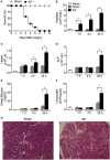
Figure 1
Figure 1
Obstruction of the biliopancreatic duct triggers severe acute pancreatitis (AP) in mice. (A) Survival of C57BL/6 mice subjected to AP (n = 31) or the sham operation (Sham, n = 16). Serum concentrations of amylase (B), lipase (C), alkaline phosphatase (ALP) (D), direct bilirubin (E), and total leukocytes (F) in the peritoneal cavity lavage were evaluated 1, 4, and 24 h after surgery in AP (n = 48), Sham (n = 27), or naïve mice (n = 27). (G) Histomorphological analysis of pancreas collected from mice 24 h after Sham or AP induction. These experiments were performed independently three times. *P < 0.05 compared with Sham mice.
Figure 2
Figure 2
Meropenem accelerates the mortality rate of mice with acute pancreatitis (AP). (A) Survival rate of mice subjected to the sham operation (Sham, n = 9), Sham mice posttreated with meropenem (Sham + MER, 100 mg/kg, i.p., 12/12 h, 3 days, n = 9), AP posttreated with saline (200 µL, i.p., 12/12 h, 3 days, n = 15), or AP posttreated with meropenem (AP + MER, 100 mg/kg, i.p., 12/12 h, 3 days, n = 25). Posttreatments started 12 h after the surgeries. *P < 0.0001 compared with the Sham groups; **P < 0.0001 compared with the AP mice. (B) Survival rate of mice subjected to the procedures described in (A), with a modification that saline and meropenem were given as a pretreatment (100 mg/kg, i.p., 12/12 h, 3 days). Surgeries were performed 12 h after the last dose of saline or meropenem (Sham n = 9, Sham + MER n = 12, AP n = 16, and AP + MER n = 31). *P < 0.0001 compared with the Sham groups; **P < 0.0001 compared with the AP mice. These experiments were performed independently three times. Serum concentrations of (C) amylase and (D) lipase of mice subjected to Sham (n = 6), Sham + MER (n = 6), AP (n = 10), and AP + MER (n = 12). Samples were collected 60 h after surgery. Serum concentrations of (E) amylase and (F) lipase in mice subjected to Sham (n = 6), MER + Sham (n = 6), AP (n = 10), and MER + AP (n = 10). Samples were collected 24 h after surgery. *P < 0.0001 compared with the Sham or naïve mice. These experiments were performed independently two times.
Figure 3
Figure 3
Meropenem pretreatment increases the colony-forming units (CFUs) in the blood of mice with acute pancreatitis (AP). The bacterial load in the (A) pancreas, (B) spleen, and (C) blood was evaluated 12 or 24 h after the surgeries in samples harvested from the pretreated mice. *P < 0.05 compared with the Sham mice. **P < 0.05 compared with the AP mice. Sham (n = 9), MER + Sham (n = 9), AP (n = 15), and MER + AP (n = 15). These experiments were performed independently three times. The pictures of the Mueller-Hinton agar plates provided in panel (C) show an example of the bacterial load in the blood samples from the AP or meropenem-pretreated AP mice 24 h after the surgeries. The pictures were converted to black and white, and the contrast of the whole picture was increased using the Adobe Photoshop 7.0 software (see original pictures in Figure S6 in Supplementary Material). (D) The CFUs that increased in the blood samples from AP or MER + AP were identified using the Vitek2 method. The results were expressed as the incidence of bacterial species in the blood (we analyzed 9 samples from a total of 15 mice). ND, not detectable.
Figure 4
Figure 4
Microbiota enrichment with Enterococcus gallinarum induces enhanced mortality of acute pancreatitis (AP) mice. (A) Experimental design for the intestinal bacterial community reduction, intestinal bacterial reconstitution, AP induction, and data analysis (mice design from https://mindthegraph.com). (B) Fresh fecal samples were collected from mice prior to the experiment (day 0) and spread on Mueller-Hinton agar plates; the results were expressed as colony-forming unit (CFU)/g of feces. Beginning on the following day (day 1) and continuing to day 15, mice were exposed to a mix of antibiotics (ABX). From day 16 to day 27, mice were gavaged daily with 100 µL (100 mg/mL) of the feces suspension or with 100 µL (1 × 108 CFU) of the E. gallinarum suspension (see Materials and Methods). Fresh feces were also collected from mice on days 5, 13, 21, and 25 and spread on Mueller-Hinton agar plates. (C) Fresh feces collected on days 21 and 25 were spread on bile esculin azide agar plates with 6 µg/mL of vancomycin, and the CFUs that grew on samples from mice treated with the E. gallinarum suspension were identified as E. gallinarum using the Vitek2 method. The results were expressed as E. gallinarum CFU/g of feces. (D) The survival rates of the Sham or AP mice that received feces or the E. gallinarum suspension (Sham n = 9, MER + Sham n = 9, AP n = 15, and MER + AP n = 15); *P < 0.0001 compared with the Sham groups; **P < 0.0001 compared with AP mice. (E) Blood samples were collected 24 h after surgery and spread on bile esculin azide agar plates with 6 µg/mL of vancomycin. CFUs grew on the samples from mice treated with the E. gallinarum suspension were identified as E. gallinarum using the Vitek2 method. The results were expressed as E. gallinarum CFU/mL of blood. (F) The survival rates of the AP mice treated intravenously (i.v.) with saline, heat-killed (dead, n = 15) E. gallinarum (1 × 108 CFU/mouse, n = 15) or live E. gallinarum (1 × 108 CFU/mouse, n = 15) and Sham mice treated i.v. with live E. gallinarum (1 × 108 CFU/mouse, n = 8); *P < 0.0001 compared with AP + dead E. gallinarum or AP + saline. The survival rates of C57BL/6 (Wt) and TLR2-deficient (TLR2−/−) mice challenged with intraperitoneal administration of (G) E. gallinarum (1 × 109/mouse, n = 15) isolated from mice or (H) E. gallinarum (1 × 109/mouse, n = 15) isolated from a septic patient. *P < 0.0001 compared with Wt E. gallinarum. **P < 0.01 compared with Wt E. gallinarum from septic patient. These experiments were performed independently three times. ND, not detectable.

Similar articles

Cited by

References

    1. Kamada N, Seo SU, Chen GY, Nunez G. Role of the gut microbiota in immunity and inflammatory disease. Nat Rev Immunol (2013) 13:321–35.10.1038/nri3430 - DOI - PubMed
    1. Levy M, Kolodziejczyk AA, Thaiss CA, Elinav E. Dysbiosis and the immune system. Nat Rev Immunol (2017) 17:219–32.10.1038/nri.2017.7 - DOI - PubMed
    1. Ayres JS, Trinidad NJ, Vance RE. Lethal inflammasome activation by a multidrug-resistant pathobiont upon antibiotic disruption of the microbiota. Nat Med (2012) 18:799–806.10.1038/nm.2729 - DOI - PMC - PubMed
    1. Ubeda C, Taur Y, Jenq RR, Equinda MJ, Son T, Samstein M, et al. Vancomycin-resistant Enterococcus domination of intestinal microbiota is enabled by antibiotic treatment in mice and precedes bloodstream invasion in humans. J Clin Invest (2010) 120:4332–41.10.1172/JCI43918 - DOI - PMC - PubMed
    1. Knoop KA, Mcdonald KG, Kulkarni DH, Newberry RD. Antibiotics promote inflammation through the translocation of native commensal colonic bacteria. Gut (2016) 65:1100–9.10.1136/gutjnl-2014-309059 - DOI - PMC - PubMed