Review
. 2018 Jan 9:8:1921.
doi: 10.3389/fimmu.2017.01921. eCollection 2017.

New Insights into the Immunobiology of Mononuclear Phagocytic Cells and Their Relevance to the Pathogenesis of Cardiovascular Diseases

Affiliations
Review

New Insights into the Immunobiology of Mononuclear Phagocytic Cells and Their Relevance to the Pathogenesis of Cardiovascular Diseases

Liliana Maria Sanmarco et al. Front Immunol. .

Abstract

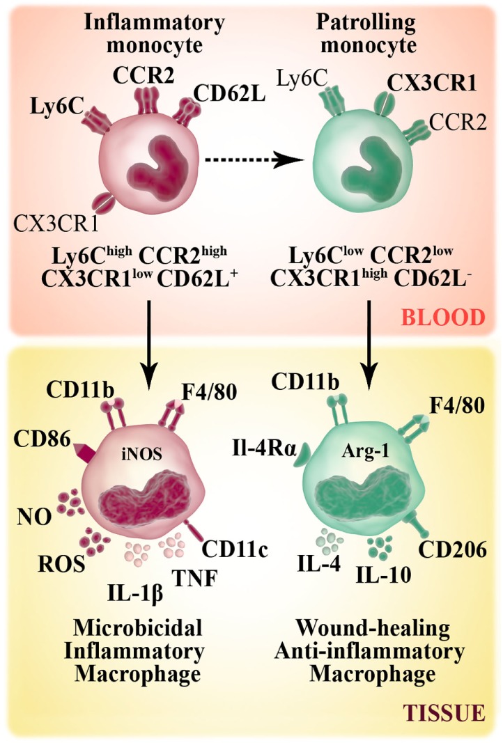
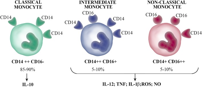
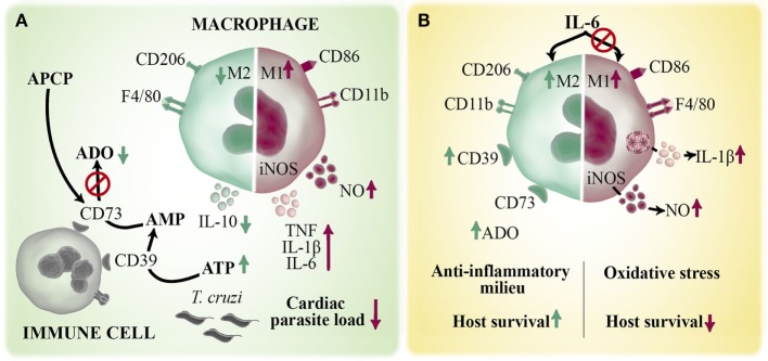
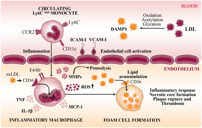
Macrophages are the primary immune cells that reside within the myocardium, suggesting that these mononuclear phagocytes are essential in the orchestration of cardiac immunity and homeostasis. Independent of the nature of the injury, the heart triggers leukocyte activation and recruitment. However, inflammation is harmful to this vital terminally differentiated organ with extremely poor regenerative capacity. As such, cardiac tissue has evolved particular strategies to increase the stress tolerance and minimize the impact of inflammation. In this sense, growing evidences show that mononuclear phagocytic cells are particularly dynamic during cardiac inflammation or infection and would actively participate in tissue repair and functional recovery. They respond to soluble mediators such as metabolites or cytokines, which play central roles in the timing of the intrinsic cardiac stress response. During myocardial infarction two distinct phases of monocyte influx have been identified. Upon infarction, the heart modulates its chemokine expression profile that sequentially and actively recruits inflammatory monocytes, first, and healing monocytes, later. In the same way, a sudden switch from inflammatory macrophages (with microbicidal effectors) toward anti-inflammatory macrophages occurs within the myocardium very shortly after infection with Trypanosoma cruzi, the causal agent of Chagas cardiomyopathy. While in sterile injury, healing response is necessary to stop tissue damage; during an intracellular infection, the anti-inflammatory milieu in infected hearts would promote microbial persistence. The balance of mononuclear phagocytic cells seems to be also dynamic in atherosclerosis influencing plaque initiation and fate. This review summarizes the participation of mononuclear phagocyte system in cardiovascular diseases, keeping in mind that the immune system evolved to promote the reestablishment of tissue homeostasis following infection/injury, and that the effects of different mediators could modulate the magnitude and quality of the immune response. The knowledge of the effects triggered by diverse mediators would serve to identify new therapeutic targets in different cardiovascular pathologies.

Keywords: Chagas disease; atherosclerosis; interleukin-6; macrophages; monocytes; oxidative stress; oxidized phospholipids; purinergic signaling.


Figures

Figure 1
Figure 1
Murine monocyte and macrophage subsets. Murine monocytes develop from a common myeloid progenitor and leave the bone marrow through the chemokine receptor CCR2. In bloodstream, circulating monocytes are phenotypically and functionally heterogeneous. Non-classical monocytes (Ly6Clow CCR2low CX3CR1high CD62L) patrol the vasculature and accumulate at low numbers in the steady state. Inflammatory or classical (Ly6Chigh CCR2high CX3CR1low CD62L+) monocytes have a relatively short-circulating lifespan and preferentially accumulate in inflammatory sites where they give rise to inflammatory M1 macrophages (F4/80+ CD11b+ CD86+ CD206). M1 macrophage subset has high microbicidal capacity due to their ability to produce inflammatory cytokines [TNF, IL-1β], reactive oxygen species (ROS) secretion and the expression of iNOS enzyme that metabolizes arginine to arginine-derived killer molecule NO. Non-classical monocytes can be recruited to tissue and differentiate to M2 macrophages (F4/80+ CD11b+ CD86 CD206+), which secrete anti-inflammatory cytokines (IL-10, IL-4) and contribute to tissue repair mechanisms.
Figure 2
Figure 2
Human circulating monocyte subsets. Human blood monocytes can be separated into three subsets according to the CD16 and CD14 expression: classical monocytes (CD14++ CD16), which represent the majority of circulating monocytes produce the anti-inflammatory IL-10 upon stimulation; intermediate monocytes (CD14++ CD16+); and non-classical monocytes (CD14+ CD16++), which secrete inflammatory cytokines such as IL-1β, IL-12, TNF, and antimicrobial molecules [nitric oxide (NO) and reactive oxygen species (ROS)].
Figure 3
Figure 3
Role of macrophages in the immune response against Trypanosoma cruzi infection. Macrophages are the main infiltrating cells arriving to the myocardium early after T. cruzi infection. Different mediators can regulate the magnitude and quality of the cardiac immune response by modulating macrophages activation. (A) ATP is released by infected/injured cells and hydrolyzed by two ectoenzymes, CD39 and CD73, to the immunoregulatory metabolite ADO. Pharmacological inhibition of CD73 activity with APCP enhances M1 over M2 macrophage phenotype and increases the local production of TNF, IL-1β, IL-6, and NO and diminished IL-10 levels. (B) IL-6 is a pleiotropic cytokine that contributes to the establishment of cardiac M2 macrophage profile during T. cruzi infection by inducing an anti-inflammatory microenvironment and augmented CD39 expression. Deficiency of IL-6 (IL6KO mice) dysregulates inflammasome activation with a consequent increases in IL1-β-induced NO production. The excessive oxidative stress and exacerbated pro-inflammatory immune response cause the lethal effect observed in infected IL6KO mice.
Figure 4
Figure 4
Role of macrophages in the pathogenesis of atherosclerosis. Disturbance of normal vascular physiology is characterized by endothelial activation which stimulates the expression of endothelial adhesion molecules (VCAM-1, ICAM-1) to favor the passage of inflammatory monocytes to the subendothelium. Modified LDL and other DAMPS are recognized by pattern-recognition receptors such as CD36 (scavenger receptor-B) in infiltrating macrophages. Thus, these innate cells are transformed into lipid-laden foam cells in the vasculature intima. The activation of NF-κB in response to ox-LDL induce the secretion of inflammatory cytokines (IL-1β, TNF), NO, MCP-1, and macrophage-derived matrix metalloproteinases (MMPs) that degrade the extracellular matrix generating a maladaptative inflammatory response and contribute to plaque formation.

References

    1. Swirski FK, Libby P, Aikawa E, Alcaide P, Luscinskas FW, Weissleder R, et al. Ly-6Chi monocytes dominate hypercholesterolemia-associated monocytosis and give rise to macrophages in atheromata. J Clin Invest (2007) 117(1):195–205. 10.1172/JCI29950 - DOI - PMC - PubMed
    1. Schulz C, Gomez Perdiguero E, Chorro L, Szabo-Rogers H, Cagnard N, Kierdorf K, et al. A lineage of myeloid cells independent of Myb and hematopoietic stem cells. Science (2012) 336(6077):86–90. 10.1126/science.1219179 - DOI - PubMed
    1. Hashimoto D, Chow A, Noizat C, Teo P, Beasley MB, Leboeuf M, et al. Tissue-resident macrophages self-maintain locally throughout adult life with minimal contribution from circulating monocytes. Immunity (2013) 38(4):792–804. 10.1016/j.immuni.2013.04.004 - DOI - PMC - PubMed
    1. Wynn TA, Chawla A, Pollard JW. Macrophage biology in development, homeostasis and disease. Nature (2013) 496(7446):445–55. 10.1038/nature12034 - DOI - PMC - PubMed
    1. Lavine KJ, Epelman S, Uchida K, Weber KJ, Nichols CG, Schilling JD, et al. Distinct macrophage lineages contribute to disparate patterns of cardiac recovery and remodeling in the neonatal and adult heart. Proc Natl Acad Sci U S A (2014) 111(45):16029–34. 10.1073/pnas.1406508111 - DOI - PMC - PubMed