. 2018 Jan;15(1):503-508.
doi: 10.3892/ol.2017.7313. Epub 2017 Oct 31.

Study on the diagnosis of small hepatocellular carcinoma caused by hepatitis B cirrhosis via multi-slice spiral CT and MRI

Affiliations

Study on the diagnosis of small hepatocellular carcinoma caused by hepatitis B cirrhosis via multi-slice spiral CT and MRI

Mei Wang et al. Oncol Lett. 2018 Jan.

Abstract

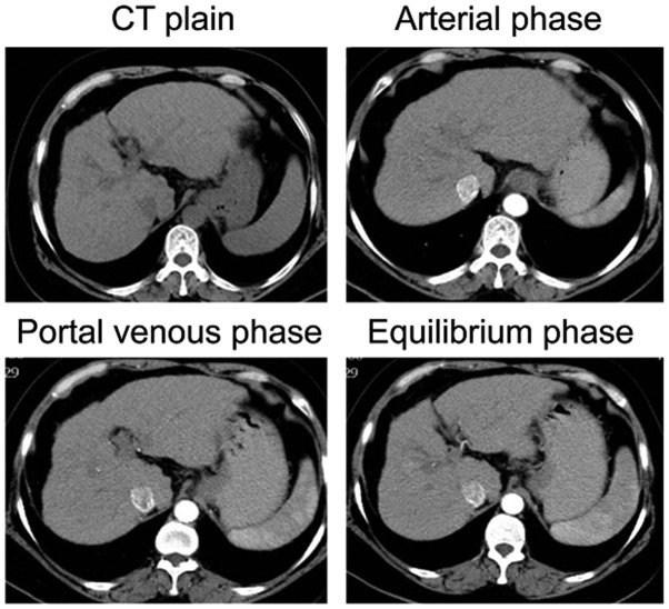
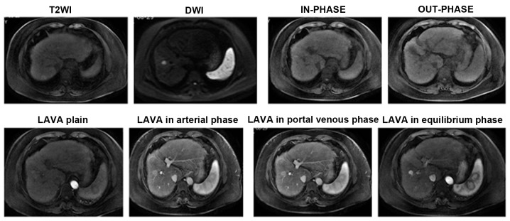
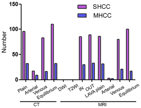
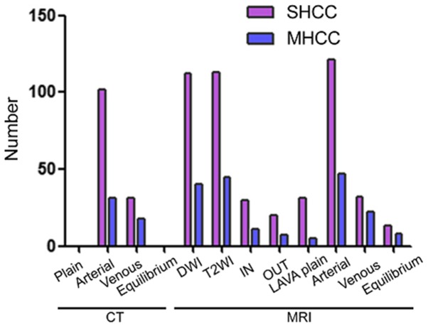
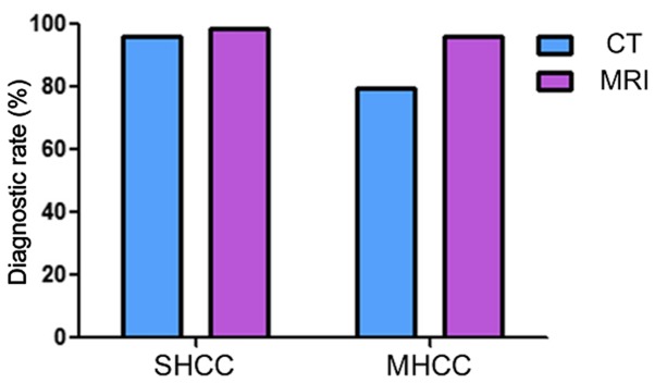
The present study compared the diagnostic accuracy of multi-slice spiral computed tomography (CT) and magnetic resonance imaging (MRI) on small hepatocellular carcinoma (SHCC) caused by hepatitis B cirrhosis. A total of 160 patients with hepatitis B cirrhosis were selected between January 2012 and April 2016, and 183 SHCC lesions were included in the present retrospective study. Patients were divided into the SHCC group (T stage) and the micro hepatocellular carcinoma (MHCC) group (T1 stage). There were a total of 129 SHCC lesions and 54 MHCC lesions identified. All patients underwent multiphasic CT and MRI imaging. The liver acquisition with volume acquisition (LAVA) technique was utilized for MRI. Furthermore, SPSS 20.0 was used for statistical analyses. LAVA in the arterial phase and CT in the arterial phase revealed significantly higher diagnostic rates for the diagnoses of 183 lesions. In addition, standard CT scan exhibited significantly reduced diagnostic rates in SHCC lesions. Results indicated that LAVA in the equilibrium phase had the lowest diagnostic rate in MHCC lesions, which was statistically significant (P<0.05). Overall, the diagnostic rate of CT (79.63%) for MHCC was significantly lower than that of MRI (96.29%) (P<0.05). However, the diagnostic rate of CT for SHCC (96.12%) was significantly higher than that for MHCC (79.63%) (P<0.05). MRI-LAVA in the arterial phase has the highest diagnostic rate for SHCC and MHCC. However, the diagnostic capability of MRI for MHCC lesions is superior to that of CT.

Keywords: diagnostic rate; magnetic resonance imaging; multi-slice spiral computed tomography; small hepatocellular carcinoma.


Figures

Figure 1.
Figure 1.
CT plain scan and images in arterial phase, portal venous phase, and equilibrium phase for SHCC patients. CT, computed tomography; SHCC, small hepatocellular carcinoma.
Figure 2.
Figure 2.
MRI examination of T2WI, DWI, IN-PHASE, OUT-PHASE, LAVA plain scan, and LAVA in arterial phase, portal venous phase, and equilibrium phase. MRI, magnetic resonance imaging; DWI, diffusion weighted imaging; T2WI, T2 weighted imaging; LAVA, liver acquisition with volume acquisition.
Figure 3.
Figure 3.
Low density in each phase of CT and low signal in each sequence of MRI of micro/small hepatocellular carcinoma lesions; except for MRI-T2WI and DWI, the distribution was different in each phase and each sequence. CT, computed tomography; MRI, magnetic resonance imaging; SHCC, small hepatocellular carcinoma; MHCC, micro hepatocellular carcinoma; DWI, diffusion weighted imaging; T2WI, T2 weighted imaging; LAVA, liver acquisition with volume acquisition.
Figure 4.
Figure 4.
Medium density in each phase of CT and equal signal in each sequence of MRI of micro/small hepatocellular carcinoma lesions; the distribution was different in 12 phases and sequences. CT, computed tomography; MRI, magnetic resonance imaging; SHCC, small hepatocellular carcinoma; MHCC, micro hepatocellular carcinoma; DWI, diffusion weighted imaging; T2WI, T2 weighted imaging; LAVA, liver acquisition with volume acquisition.
Figure 5.
Figure 5.
High density in each phase of CT and high signal in each sequence of MRI of micro/small hepatocellular carcinoma lesions; except plain scan CT and in equilibrium phase CT, the distribution was different in each phase and each sequence. CT, computed tomography; MRI, magnetic resonance imaging; SHCC, small hepatocellular carcinoma; MHCC, micro hepatocellular carcinoma; DWI, diffusion weighted imaging; T2WI, T2 weighted imaging; LAVA, liver acquisition with volume acquisition.
Figure 6.
Figure 6.
Comparisons of diagnostic rates of multi-slice spiral CT and MRI for micro/small hepatocellular carcinoma lesions; the differences in diagnostic rate for SHCC between CT and MRI were not significant (P=0.4432), and the diagnostic rate of CT for MHCC was significantly lower than that of MRI (P=0.0007). CT, computed tomography; MRI, magnetic resonance imaging; SHCC, small hepatocellular carcinoma; MHCC, micro hepatocellular carcinoma.

Similar articles

Cited by

References

    1. Fitzmorris P, Shoreibah M, Anand BS, Singal AK. Management of hepatocellular carcinoma. J Cancer Res Clin Oncol. 2015;141:861–876. doi: 10.1007/s00432-014-1806-0. - DOI - PMC - PubMed
    1. Bruix J, Gores GJ, Mazzaferro V. Hepatocellular carcinoma: Clinical frontiers and perspectives. Gut. 2014;63:844–855. doi: 10.1136/gutjnl-2013-306627. - DOI - PMC - PubMed
    1. Fernández-Rodríguez CM, Gutiérrez-García ML. Prevention of hepatocellular carcinoma in patients with chronic hepatitis B. World J Gastrointest Pharmacol Ther. 2014;5:175–182. doi: 10.4292/wjgpt.v5.i3.175. - DOI - PMC - PubMed
    1. Hartke J, Johnson M, Ghabril M. The diagnosis and treatment of hepatocellular carcinoma. Semin Diagn Pathol. 2017;34:153–159. doi: 10.1053/j.semdp.2016.12.011. - DOI - PubMed
    1. Banaudha KK, Verma M. Epigenetic biomarkers in liver cancer. Methods Mol Biol. 2015;1238:65–76. doi: 10.1007/978-1-4939-1804-1_4. - DOI - PubMed