

Uterine artery Doppler velocimetry in hypertensive disorder of pregnancy in Nigeria
- PMID: 29375900
- PMCID: PMC5769665
- DOI: 10.15557/JoU.2017.0037
Uterine artery Doppler velocimetry in hypertensive disorder of pregnancy in Nigeria
Abstract
Aim of the study: To evaluate the value of uterine artery Doppler indices and waveform pattern in predicting fetuses at risk for intrauterine growth restriction in hypertensive disorders of pregnancy.
Materials and methods: This was a prospective cross-sectional study including 80 pregnant subjects with hypertensive disorders of pregnancy and two control groups. Uterine artery Doppler sonography was performed in all study participants. Uterine artery Doppler indices across the groups were compared using the analysis of variance (ANOVA) while the presence of prediastolic notch was analyzed with the Chi Square test.
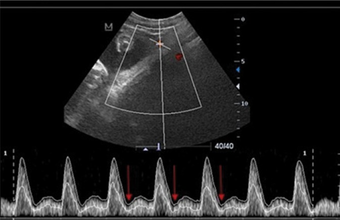
Results: For the hypertensive disorders of pregnancy group, resistivity index > 0.66 had a sensitivity of 50.0%, specificity of 69.1% and a positive predictive value of 22.2% for predicting intrauterine growth restriction. The odds ratio was 2.2 with a 95% confidence interval of 0.6-7.8. The presence of prediastolic notching had a sensitivity of 100.0%, specificity of 96.0% and a positive predictive value of 80.0% for predicting intrauterine growth restriction. The odds ratio was 22.7 with a 95% confidence interval of 7.5-68.5.
Conclusion: Uterine artery Doppler sonography is useful for predicting fetuses at risk for intrauterine growth restriction in hypertensive disorder of pregnancy. Prediastolic notching is more sensitive and more specific than uterine artery resistivity index in predicting fetuses at risk of intrauterine growth restriction in established hypertensive disorder of pregnancy.
Keywords: Doppler sonography; hypertensive disorder of pregnancy; intrauterine growth restriction; uterine artery.
Figures

Similar articles
-
Uterine artery Doppler velocimetry in the detection of adverse obstetric outcomes in women with unexplained elevated maternal serum alpha-fetoprotein levels.Am J Obstet Gynecol. 1995 Oct;173(4):1115-9. doi: 10.1016/0002-9378(95)91336-x. Am J Obstet Gynecol. 1995. PMID: 7485303 Clinical Trial.
-
Doppler assessment of the uterine and uteroplacental circulation in the second trimester in pregnancies at high risk for pre-eclampsia and/or intrauterine growth retardation: comparison and correlation between different Doppler parameters.Ultrasound Obstet Gynecol. 1997 May;9(5):330-8. doi: 10.1046/j.1469-0705.1997.09050330.x. Ultrasound Obstet Gynecol. 1997. PMID: 9201877 Clinical Trial.
-
Cell-free fetal DNA concentration in plasma of patients with abnormal uterine artery Doppler waveform and intrauterine growth restriction--a pilot study.Prenat Diagn. 2003 May;23(5):367-71. doi: 10.1002/pd.596. Prenat Diagn. 2003. PMID: 12749031
-
First-trimester uterine artery Doppler and adverse pregnancy outcome: a meta-analysis involving 55,974 women.Ultrasound Obstet Gynecol. 2014 May;43(5):500-7. doi: 10.1002/uog.13275. Epub 2014 Apr 4. Ultrasound Obstet Gynecol. 2014. PMID: 24339044 Review.
-
Third-trimester uterine artery Doppler for prediction of adverse outcome in late small-for-gestational-age fetuses: systematic review and meta-analysis.Ultrasound Obstet Gynecol. 2020 May;55(5):575-585. doi: 10.1002/uog.21940. Ultrasound Obstet Gynecol. 2020. PMID: 31785172
Cited by
-
Flow-mediated Dilation of the Brachial Artery in Women with Hypertensive Disorders of Pregnancy.J Med Ultrasound. 2023 Oct 6;32(1):48-54. doi: 10.4103/jmu.jmu_10_23. eCollection 2024 Jan-Mar. J Med Ultrasound. 2023. PMID: 38665342 Free PMC article.
-
Differences in Clinical Characteristics and Therapy of Neonatal Acute Respiratory Distress Syndrome (ARDS) and Respiratory Distress Syndrome (RDS): A Retrospective Analysis of 925 Cases.Med Sci Monit. 2019 Jul 6;25:4992-4998. doi: 10.12659/MSM.915213. Med Sci Monit. 2019. PMID: 31278248 Free PMC article.
-
Fetal Gestational Age Estimation Using Ultrasonic Transverse Cerebellar Diameter in a Sub-Saharan African Population.J Med Ultrasound. 2023 Jun 2;32(1):41-47. doi: 10.4103/jmu.jmu_116_22. eCollection 2024 Jan-Mar. J Med Ultrasound. 2023. PMID: 38665343 Free PMC article.
-
Association between Endothelial Dysfunction, Biomarkers of Renal Function, and Disease Severity in Sickle Cell Disease.Kidney360. 2020 Jan 31;1(2):79-85. doi: 10.34067/KID.0000142019. eCollection 2020 Feb 27. Kidney360. 2020. PMID: 35372907 Free PMC article.
-
Comparative Study of the Umbilical Artery Doppler Indices of Healthy and Growth-Restricted Foetuses in Lagos.J West Afr Coll Surg. 2022 Apr-Jun;12(2):63-69. doi: 10.4103/jwas.jwas_63_22. Epub 2022 Aug 27. J West Afr Coll Surg. 2022. PMID: 36213799 Free PMC article.
References
-
- Ebeigbe P, Aziken M. Early onset pregnancy-induced hypertension/eclampsia in Benin City, Nigeria. Niger J Clin Pract. 2010;13:388–393. - PubMed
-
- Khong TY, De Wolf F, Robertson WB, Brosens I. Inadequate maternal vascular response to placentation in pregnancies complicated by pre-eclampsia and by small-for-gestational age infants. Br J Obstet Gynaecol. 1986;93:1049–1059. - PubMed
-
- Program NHBPE Report of the national high blood pressure education program working group on high blood pressure in pregnancy. Am J Obstet Gynecol. 2000;183(1):S1–S22. - PubMed
-
- Lakhkar BN, Ahamed SA. Doppler velocimetry of uterine and umbilical arteries during pregnancy. Indian J Radiol Imaging. 1999;9:119–125.
LinkOut - more resources
Full Text Sources
Other Literature Sources
Research Materials