

Cognitive clinico-radiological paradox in early stages of multiple sclerosis
- PMID: 29376094
- PMCID: PMC5771324
- DOI: 10.1002/acn3.512
Cognitive clinico-radiological paradox in early stages of multiple sclerosis
Abstract
Objective: To investigate whether the strength of the association between magnetic resonance imaging (MRI) metrics and cognitive outcomes differs between various multiple sclerosis subpopulations.
Methods: A total of 1052 patients were included in this large cross-sectional study. Brain MRI (T1 and T2 lesion volume and brain parenchymal fraction) and neuropsychological assessment (Brief International Cognitive Assessment for Multiple Sclerosis and Paced Auditory Serial Addition Test) were performed.
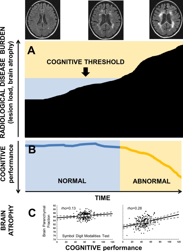
Results: Weak correlations between cognitive domains and MRI measures were observed in younger patients (age≤30 years; absolute Spearman's rho = 0.05-0.21), with short disease duration (<2 years; rho = 0.01-0.21), low Expanded Disability Status Scale [EDSS] (≤1.5; rho = 0.08-0.18), low T2 lesion volume (lowest quartile; <0.59 mL; rho = 0.01-0.20), and high brain parenchymal fraction (highest quartile; >86.66; rho = 0.01-0.16). Stronger correlations between cognitive domains and MRI measures were observed in older patients (age>50 years; rho = 0.24-0.50), with longer disease duration (>15 years; rho = 0.26-0.53), higher EDSS (≥5.0; rho = 0.23-0.39), greater T2 lesion volume (highest quartile; >5.33 mL; rho = 0.16-0.32), and lower brain parenchymal fraction (lowest quartile; <83.71; rho = 0.13-0.46). The majority of these observed results were confirmed by significant interactions (P ≤ 0.01) using continuous variables.
Interpretation: The association between structural brain damage and functional cognitive impairment is substantially weaker in multiple sclerosis patients with a low disease burden. Therefore, disease stage should be taken into consideration when interpreting associations between structural and cognitive measures in clinical trials, research studies, and clinical practice.
Figures

References
-
- Benedict RH, Zivadinov R. Risk factors for and management of cognitive dysfunction in multiple sclerosis. Nat Rev Neurol 2011;7:332–342. - PubMed
-
- Uher T, Blahova‐Dusankova J, Horakova D, et al. Longitudinal MRI and neuropsychological assessment of patients with clinically isolated syndrome. J Neurol 2014;261:1735–1744. - PubMed
-
- Rocca MA, Amato MP, De Stefano N, et al. Clinical and imaging assessment of cognitive dysfunction in multiple sclerosis. Lancet Neurol 2015;14:302–317. - PubMed
-
- Uher T, Vaneckova M, Sobisek L, et al. Combining clinical and magnetic resonance imaging markers enhances prediction of 12‐year disability in multiple sclerosis. Mult Scler. 2017;23:51–61. - PubMed
-
- Morrow SA, Drake A, Zivadinov R, et al. Predicting loss of employment over three years in multiple sclerosis: clinically meaningful cognitive decline. Clin Neuropsychol. 2010;24:1131–1145. - PubMed
LinkOut - more resources
Full Text Sources
Other Literature Sources
