. 2018 Jan;5(1):011019.
doi: 10.1117/1.NPh.5.1.011019. Epub 2018 Jan 19.

Are ventrolateral and dorsolateral prefrontal cortices involved in the computerized Corsi block-tapping test execution? An fNIRS study

Affiliations

Are ventrolateral and dorsolateral prefrontal cortices involved in the computerized Corsi block-tapping test execution? An fNIRS study

Stefania Lancia et al. Neurophotonics. 2018 Jan.

Abstract

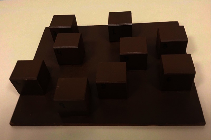
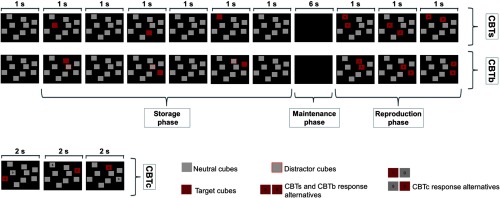
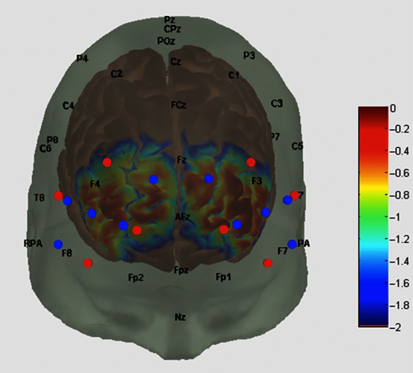
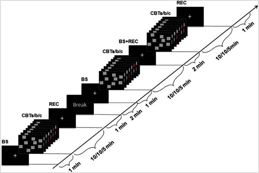
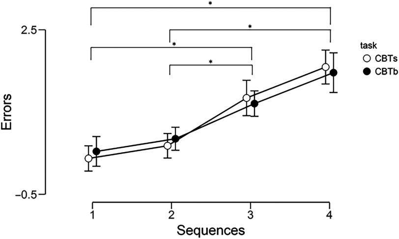
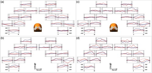
The Corsi block-tapping test (CBT) is an old neuropsychological test that, requiring the storage and the reproduction of spatial locations, assesses spatial working memory (WM). Despite its wide use in clinical practice, the specific contribution of prefrontal cortex (PFC) subregions during CBT execution has not been clarified yet. Considering the importance of spatial WM in daily life and the well-known role of ventrolateral-PFC/dorsolateral-PFC (VLPFC/DLPFC) in WM processes, the present study was aimed at investigating, by a 20-channel functional near-infrared spectroscopy (fNIRS) system (including four short-separation channels), the hemodynamic response of the VLPFC/DLPFC during a computerized version of the CBT. Thirty-nine university students were asked to perform CBT standard version (CBTs), block-suppression CBT (CBTb), and control task (CBTc). A VLPFC activation during CBTs and a DLPFC activation during CBTb were hypothesized. The results of the Bayesian analysis have not shown a delineated specific activation of VLPFC/DLPFC during either CBTs or CBTb. These results together with the related ones obtained by others using fMRI are not sufficient to definitively state the role of the PFC subregions during CBT execution. The adoption of high-density diffuse optical tomography would be helpful in further exploration of the PFC involvement in spatial WM tasks.

Keywords: Corsi block-tapping test; dorsolateral prefrontal cortex; fNIRS; ventrolateral prefrontal cortex; working memory.


Figures

Fig. 1
Fig. 1
The standard version of the Corsi block-tapping board. The base of wood board was 29×24  cm and the 9 cubes measured 4 cm.
Fig. 2
Fig. 2
Representation of a three-cube sequence during the CBTs, the CBTb, and the CBTc. During CBTs and CBTb, each target cube was usually presented on the computer screen for 1 s. The interval between target cube presentations was 1 s, during which a no-target cube was shown on the screen. The storage phase was followed by a black screen lasting 6 s. The presentation time on the screen during CBTc was 2 s. Storage and reproduction phases of the CBTs and the CBTb are illustrated. In the reproduction phases of CBTs and CBTb and during the CBTc, some cubes were labeled with “S” (“sinistra” in Italian, “left” in English) for the cube that was located more left in relation to the second one, which was labeled with “D” (“destra” in Italian, “right” in English) located more right (see Corsi block-tapping test-computerized version section for more details).
Fig. 3
Fig. 3
Map of sensitivity profile of the 16 measurement points and 4 short-separation channels. The probe layout was registered and anchored on the digital brain adult atlas (Colin27), and sensitivity profile of the 16 measurement points and 4 short-separation channels is represented. Red and blue circles represent the illuminators and detectors, respectively. The color scale depicts the sensitivity profile from 2 (low sensitivity) to 0 (high sensitivity). See Sec. 2 for details.
Fig. 4
Fig. 4
Schematic representation of the protocol. BS, baseline period; CBTs/b/c, Corsi block-tapping test standard/block-suppression/control task; REC, recovery period.
Fig. 5
Fig. 5
Results of the posthoc comparison for the performance among the four sequences. Results are shown as mean errors per sequence length with 95% credible intervals. *, BF10>100.
Fig. 6
Fig. 6
Grand average (n=39) of the PFC O2Hb (solid lines) and HHb (dotted lines) changes in response to the standard Corsi block-tapping test (red) and the Corsi block-tapping test block-suppression (blue) during the four sequence length. (a) Three-cube sequence; (b) four-cube sequence; (c) five-cube sequence; and (d) six-cube sequence. The layout of the 16 fNIRS measurement points over the right (numbers from 1 to 8) and left (numbers from 9 to 16) hemisphere is graphically represented. The vertical solid lines limit the duration of the task execution of each sequence.

Similar articles

Cited by

References

    1. Baddeley A. D., Hitch G., Bower G. A., “Working memory,” in Recent Advances in Learning and Motivation, Academic Press, New York: (1974).
    1. Baddeley A. D., “Working memory and language: an overview,” J. Commun. Disord. 36(3), 189–208 (2003).10.1016/S0021-9924(03)00019-4 - DOI - PubMed
    1. Collette F., et al. , “Exploration of the neural substrates of executive functioning by functional neuroimaging,” Neuroscience 139(1), 209–221 (2006).10.1016/j.neuroscience.2005.05.035 - DOI - PubMed
    1. Hartley A. A., Speer N. K., “Locating and fractionating working memory using functional neuroimaging: storage, maintenance, and executive functions,” Microsc. Res. Technol. 51(1), 45–53 (2000).10.1002/1097-0029(20001001)51:1<45::AID-JEMT5>3.0.CO;2-O - DOI - PubMed
    1. Lara A. H., Wallis J. D., “The role of prefrontal cortex in working memory: a mini review,” Front. Syst. Neurosci. 9, 173 (2015).10.3389/fnsys.2015.00173 - DOI - PMC - PubMed