

Bevacizumab Injection in Patients with Neovascular Age-Related Macular Degeneration Increases Angiogenic Biomarkers
- PMID: 29376143
- PMCID: PMC5783314
- DOI: 10.1016/j.oret.2017.04.004
Bevacizumab Injection in Patients with Neovascular Age-Related Macular Degeneration Increases Angiogenic Biomarkers
Abstract
Purpose: To evaluate the expression of 19 angiogenic biomarkers in the aqueous humor before and after intravitreal bevacizumab injection (IVB) in eyes with neovascular age-related macular degeneration (AMD).
Design: Prospective, noncomparative, interventional case series.
Participants: Twenty-three eyes of 23 treatment-naïve patients with choroidal neovascularization (CNV) secondary to neovascular AMD.
Methods: Eyes were diagnosed with CNV secondary to neovascular AMD and were treated with 3 monthly IVBs. Aqueous humor samples were obtained by anterior chamber paracentesis at baseline and immediately before each intravitreal bevacizumab injection.
Main outcome measures: Aqueous humor levels of 19 angiogenic biomarkers (angiopoietin 2, bone morphogenetic protein 9 [BMP-9], epidermal growth factor [EGF], endoglin, endothelin 1, fibroblast growth factor [FGF]-1 and FGF-2, follistatin, granulocyte colony-stimulating factor [GCSF], heparin-binding EGF-like growth factor [HB-EGF], hepatocyte growth factor [HGF], interleukin 8, leptin, placental growth factor [PLGF], vascular endothelial growth factor [VEGF]-A, VEGF-C, VEGF-D, and tissue inhibitor of metalloproteinases [TIMP]-1 and TIMP-2) were measured. Best-corrected visual acuity (BCVA), spectral-domain OCT parameters, and intraocular pressure also were evaluated.
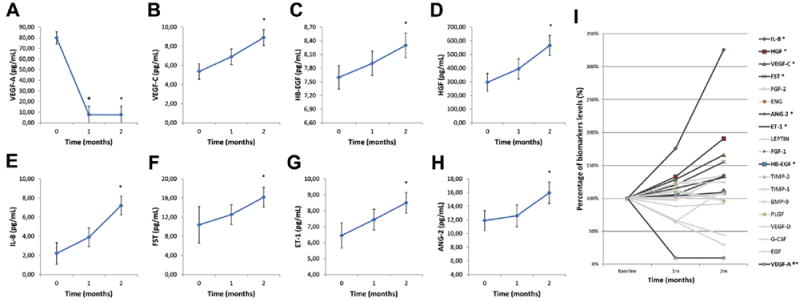
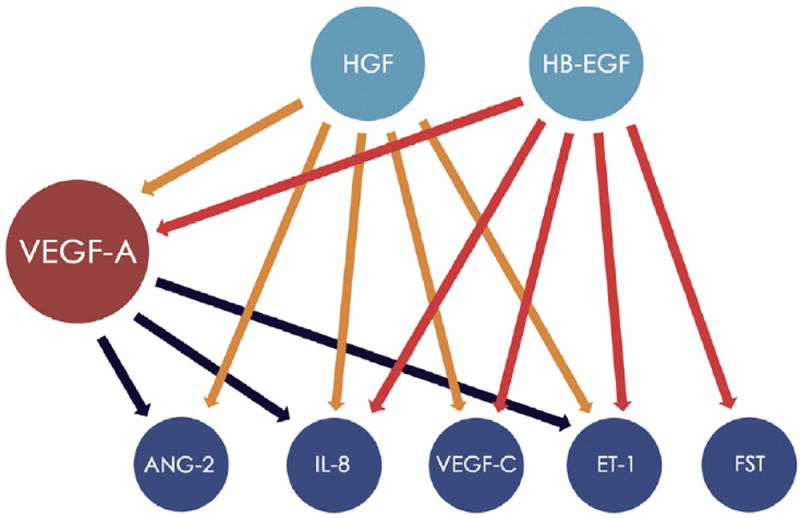
Results: Baseline aqueous VEGF-A expression was elevated in all study eyes before treatment initiation. A statistically significant decrease of VEGF-A was observed at the 1- and 2-month follow-ups. A statistically significant increased concentration was observed in 7 biomarkers: VEGF-C, angiopoietin 2, endothelin 1, follistatin, HB-EGF, HGF, and interleukin 8. The other 11 study biomarker levels (VEGF-D, BMP-9, EGF, endoglin, FGF-1, FGF-2, GCSF, leptin, PLGF, TIMP-1, and TIMP-2) did not show any significant difference during follow-up. The BCVA statistically improved significantly at 2 months. Spectral-domain OCT parameters improved significantly at all follow-ups. Mean intraocular pressure values were not statistically different during the study period.
Conclusions: Despite a decrease in VEGF-A, the aqueous levels of VEGF-C, angiopoietin 2, endothelin 1, follistatin, HB-EGF, HGF, and interleukin 8 increased significantly after intravitreal injection of bevacizumab. These upregulated angiogenic biomarkers may represent new therapeutic targets in exudative AMD.
Figures
References
-
- Carmeliet P. Angiogenesis in health and disease. Nat Med. 2003;9:653–660. - PubMed
-
- Kijlstra A, La Heij E, Hendrikse F. Immunological factors in the pathogenesis and treatment of age-related macular degeneration. Ocul Immunol Inflamm. 2005;13:3–11. - PubMed
-
- Group EDPR. Causes and prevalence of visual impairment among adults in the United States. Arch Ophthalmol. 2004;122:477–485. - PubMed
-
- Lopez PF, Sippy BD, Lambert HM, et al. Transdifferentiated retinal pigment epithelial cells are immunoreactive for vascular endothelial growth factor in surgically excised age-related macular degeneration-related choroidal neovascular membranes. Invest Ophthalmol Vis Sci. 1996;37:855–868. - PubMed
-
- Heier JS, Brown DM, Chong V, et al. Intravitreal aflibercept (VEGF trap-eye) in wet age-related macular degeneration. Ophthalmology. 2012;119:2537–2548. - PubMed
Grants and funding
LinkOut - more resources
Full Text Sources
Other Literature Sources
Medical
Research Materials
Miscellaneous