

Joint Craniomaxillofacial Bone Segmentation and Landmark Digitization by Context-Guided Fully Convolutional Networks
- PMID: 29376150
- PMCID: PMC5786437
- DOI: 10.1007/978-3-319-66185-8_81
Joint Craniomaxillofacial Bone Segmentation and Landmark Digitization by Context-Guided Fully Convolutional Networks
Abstract
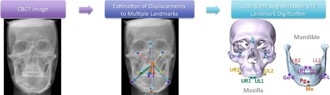
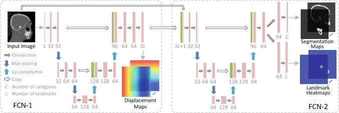
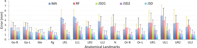
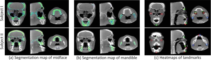
Generating accurate 3D models from cone-beam computed tomography (CBCT) images is an important step in developing treatment plans for patients with craniomaxillofacial (CMF) deformities. This process often involves bone segmentation and landmark digitization. Since anatomical landmarks generally lie on the boundaries of segmented bone regions, the tasks of bone segmentation and landmark digitization could be highly correlated. However, most existing methods simply treat them as two standalone tasks, without considering their inherent association. In addition, these methods usually ignore the spatial context information (i.e., displacements from voxels to landmarks) in CBCT images. To this end, we propose a context-guided fully convolutional network (FCN) for joint bone segmentation and landmark digitization. Specifically, we first train an FCN to learn the displacement maps to capture the spatial context information in CBCT images. Using the learned displacement maps as guidance information, we further develop a multi-task FCN to jointly perform bone segmentation and landmark digitization. Our method has been evaluated on 107 subjects from two centers, and the experimental results show that our method is superior to the state-of-the-art methods in both bone segmentation and landmark digitization.
Figures
References
-
- Cheng E, Chen J, Yang J, Deng H, Wu Y, Megalooikonomou V, Gable B, Ling H. 2011 Annual International Conference of the IEEE Engineering in Medicine and Biology Society. EMBC; 2011. Automatic dent-landmark detection in 3-D CBCT dental volumes; pp. 6204–6207. - PubMed
-
- Ronneberger O, Fischer P, Brox T. U-Net: convolutional networks for biomedical image segmentation. In: Navab N, Hornegger J, Wells WM, Frangi AF, editors. MICCAI 2015, LNCS. Vol. 9351. Springer; Cham: 2015. pp. 234–241. - DOI
Publication types
MeSH terms
Grants and funding
LinkOut - more resources
Full Text Sources
Other Literature Sources
Medical