Review
. 2018 Jul;91(1088):20170337.
doi: 10.1259/bjr.20170337. Epub 2018 Mar 2.

Paraspinal arteriovenous fistula: Stuttgart classification based on experience and a review of the literature

Affiliations
Review

Paraspinal arteriovenous fistula: Stuttgart classification based on experience and a review of the literature

C M Wendl et al. Br J Radiol. 2018 Jul.

Abstract

The term "paraspinal arteriovenous shunts" (PAVSs) summarizes an inhomogeneous variety of rare vascular disorders. PAVSs have been observed as congenital or acquired lesions. The clinical course of PAVSs may be asymptomatic or present with life-threatening symptoms. Based on a collection of individual cases from three institutions and a literature evaluation, we propose the following classification: PAVSs that are part of a genetic syndrome are separated from "isolated" PAVSs. Isolated PAVSs are subdivided into "acquired", "traumatic" and "congenital" without an identifiable genetic hereditary disorder. The subgroups are differentiated by the route of venous drainage, being exclusively extraspinal or involving intraspinal veins. PAVSs associated to a genetic syndrome may either have a metameric link or occur together with a systemic genetic disorder. Again extra-vs intraspinal venous drainage is differentiated. The indication for treatment is based on individual circumstances (e.g. myelon compression, vascular bruit, high volume output cardiac failure). Most PAVSs can be treated by endovascular means using detachable coils, liquid embolic agents or stents and derivates.


Figures

Figure 1.
Figure 1.
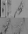
Time-of-flight MRA of a 62-year-old patient presenting with a pulsatile tinnitus of the left ear, dizziness and no history of trauma, showing arterialization of the left transverse sinus (a). Digital subtraction angiography confirms two different arteriovenous shunts, an arteriovenous shunt of the skull base [(b), injection of the ascending pharyngeal artery, black arrow] and a vertebrovertebral shunt (white arrow) originating from the right vertebral artery, the latter draining via paraspinal venous plexuses in the ipsilateral internal jugular vein (c, d). Both shunts were not treated yet, because the patient refused any kind of therapy. MRA, MR angiography.
Figure 2.
Figure 2.
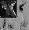
A 78-year-old female patient suffering from progressive paraparesis. The sagittal T2 weighted images (a, b) show intramedullary oedema and serpingineous leptomeningeal veins indicating a spinal AVS. The contrast-enhanced MR angiography (c) reveals a primary shunt into the epidural and paraspinal venous plexuses. DSA of the third lumbar radicular artery on the left confirms a paraspinal AVS that drains primarily into the segmental epidural spinal venous plexus (d) and into a leptomeningeal vein of the longitudinal venous drainage of the spinal cord (e). AVS, arteriovenous shunt; DSA, digital subtraction angiography.
Figure 3.
Figure 3.
Large calibre arteriovenous shunt from the descending aorta to the vena cava inferior in a 7-year-old boy with an audible bruit on his back. Venous congestion of the para-and intraspinal veins (a, b). The arteriovenous connection was occluded with coils in 2012 (c). A follow-up DSA, 4 years later confirmed the persistent occlusion of the paraspinal arteriovenous shunt (d). DSA, digital subtraction angiography.
Figure 4.
Figure 4.
A 44-year-old male patient with Klippel–Trénaunay syndrome. He presented with progressive paraparesis due to a medullary arteriovenous malformation, supplied by the anterior spinal artery, which carries a fusiform aneurysm (a). The supply of the AV-malformation and the aneurysm were occluded with coils in order to eliminate the associated bleeding risk (b), achieving a significant reduction of the arteriovenous shunt. While the clinical symptoms were caused by the spinal AVM, injection of the right common iliac artery shows an asymptomatic paraspinal arteriovenous shunt with paraspinal venous drainage (c–e). This AV-shunt remains asymptomatic and, therefore untreated. The condition of the patient stabilized, he walks with a cane. AVM, arteriovenous malformation.
Figure 5.
Figure 5.
A 50-year-old female patient with neurofibromatosis. Clumsiness of her right hand and a suspected intraspinal neurofibroma (a) led to a surgical exploration of her cervical spine. During this operation, no neurofibroma but intraspinal arterialized veins were found. The injection of the right vertebral artery showed several fusiform dilatations of this artery and an arteriovenous shunt to the para-and intraspinal veins (b, c). Both, the aneurysms and the arteriovenous shunts were occluded with detachable coils (d). Follow-up MRI, 6 months later confirmed the complete disappearance of the intraspinal veins (e).

References

    1. Kendall BE, Logue V. Spinal epidural angiomatous malformations draining into intrathecal veins. Neuroradiology 1977; 13: 181–9. doi: 10.1007/BF00344211 - DOI - PubMed
    1. Foix CH, Alajouanine T. La myélite nécrotique subaigue. Rev Neurol 1926; 2: 1–42.
    1. Iizuka Y, Suzuki M, Suzuki K, Shimoji K, Komura S. High-flow paraspinal osseous epidural arteriovenous fistula. A case report. Neuroradiol J 2008; 21: 433–9. doi: 10.1177/197140090802100322 - DOI - PubMed
    1. Rodesch G, Lasjaunias P. Spinal cord arteriovenous shunts: from imaging to management. Eur J Radiol 2003; 46: 221–32. doi: 10.1016/S0720-048X(03)00093-7 - DOI - PubMed
    1. Spetzler RF, Detwiler PW, Riina HA, Porter RW. Modified classification of spinal cord vascular lesions. J Neurosurg 2002; 96(2 Suppl): 145–56. doi: 10.3171/spi.2002.96.2.0145 - DOI - PubMed