

Comment
doi: 10.7554/eLife.34396.
Portraits of a pressure sensor
Affiliations
- PMID: 29376828
- PMCID: PMC5788499
- DOI: 10.7554/eLife.34396
Item in Clipboard
Comment
Portraits of a pressure sensor
Elife.
.
Abstract
Near atomic-resolution structures have provided insights into the mechanisms by which the Piezo1 ion channel senses and responds to mechanical stimuli.
Keywords: biophysics; cryoEM; human; mechanosensitivity; mouse; neuroscience; piezo ion channels; somatosensation; structural biology; vascular system.
Conflict of interest statement
AC, MS No competing interests declared
Figures
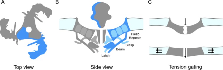
(A) The schematic structure of Piezo1 viewed from above, showing the three 'propeller blades' surrounding a central pore. A single propeller blade is highlighted in blue. (B) A side view of the structure of Piezo1 as revealed by cryo-electron microscopy, with a single propeller blade highlighted in dark and medium blue: the interior of the cell is at the bottom of the figure. Each propeller blade contains at least six piezo-repeats, but only the three nearest to the central pore are shown (medium blue). Each propeller blade also includes a 'beam' domain (dark blue) that is parallel to the cell membrane, and a structure called the 'latch' (dark blue) that is in contact with the intracellular ends of the inner helices (dark grey) that form the central pore. Each propeller also contains a 'clasp' domain (medium blue): this domain interacts further from the pore, but its structure has not been determined yet. (C) When the cell is not submitted to pressure, Piezo1 bends the membrane to make a dome-like structure pointing inside the cell, and the channel is closed. When the membrane is stretched the complex flattens out, opening the channel.
Comment on
-
Structure-based membrane dome mechanism for Piezo mechanosensitivity.Elife. 2017 Dec 12;6:e33660. doi: 10.7554/eLife.33660. Elife. 2017. PMID: 29231809 Free PMC article.
References
-
- Chesler AT, Szczot M, Bharucha-Goebel D, Čeko M, Donkervoort S, Laubacher C, Hayes LH, Alter K, Zampieri C, Stanley C, Innes AM, Mah JK, Grosmann CM, Bradley N, Nguyen D, Foley AR, Le Pichon CE, Bönnemann CG. The role of PIEZO2 in human mechanosensation. The New England Journal of Medicine. 2016;375:1355–1364. doi: 10.1056/NEJMoa1602812. - DOI - PMC - PubMed
Publication types
MeSH terms
Substances
LinkOut - more resources
Full Text Sources
Other Literature Sources
Molecular Biology Databases
