. 2018 Mar 1;128(3):960-969.
doi: 10.1172/JCI97007. Epub 2018 Jan 29.

Hypothalamic loss of Snord116 recapitulates the hyperphagia of Prader-Willi syndrome

Affiliations

Hypothalamic loss of Snord116 recapitulates the hyperphagia of Prader-Willi syndrome

Joseph Polex-Wolf et al. J Clin Invest. .

Abstract

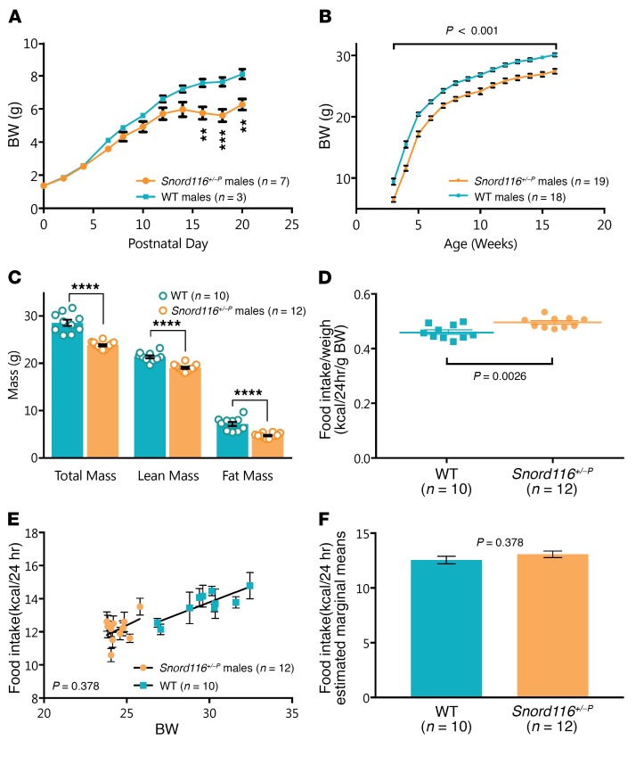
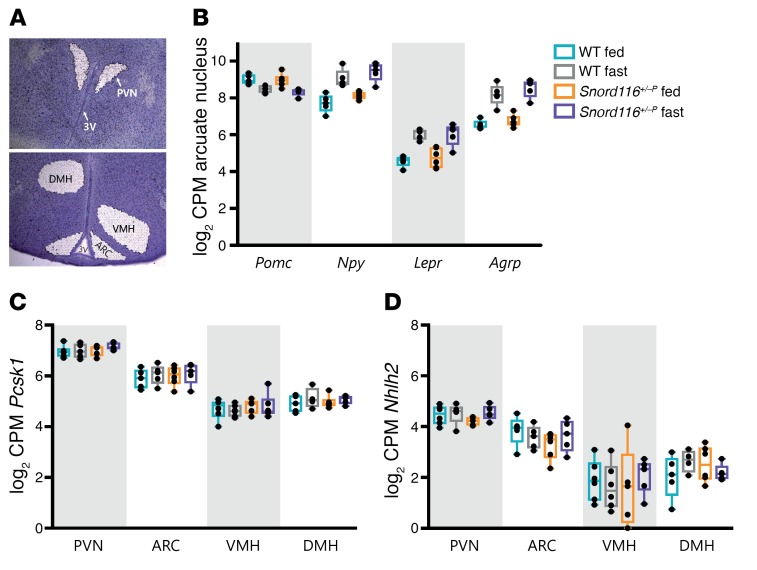
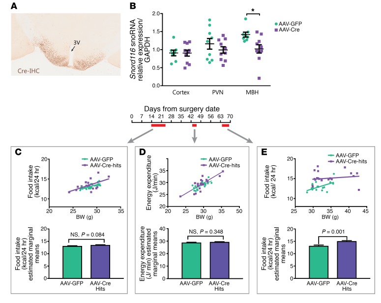
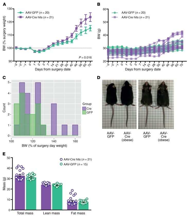
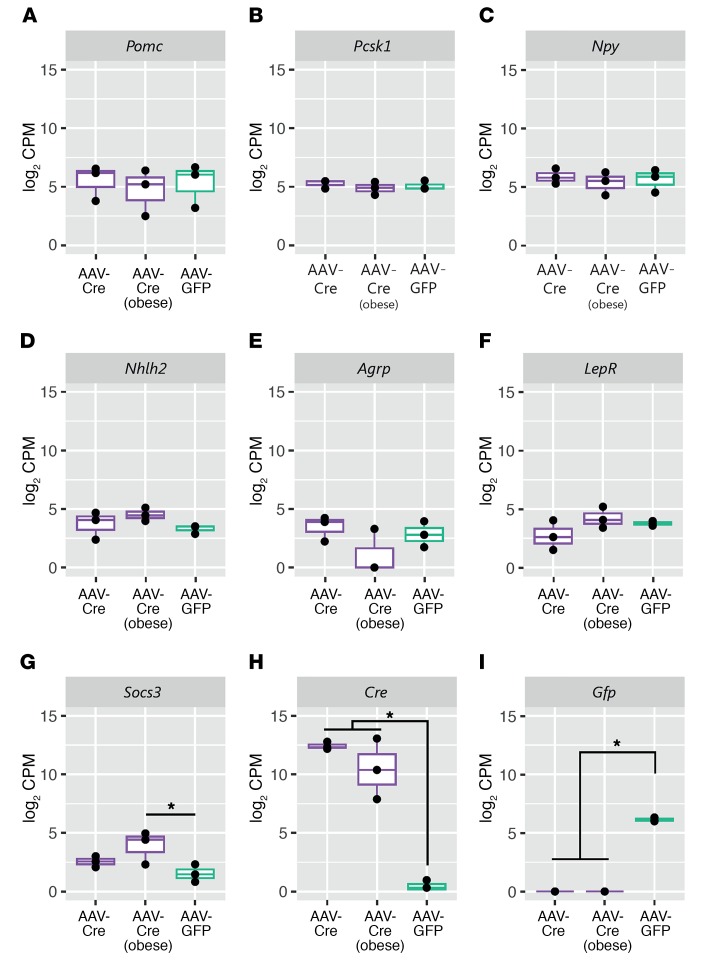
Profound hyperphagia is a major disabling feature of Prader-Willi syndrome (PWS). Characterization of the mechanisms that underlie PWS-associated hyperphagia has been slowed by the paucity of animal models with increased food intake or obesity. Mice with a microdeletion encompassing the Snord116 cluster of noncoding RNAs encoded within the Prader-Willi minimal deletion critical region have previously been reported to show growth retardation and hyperphagia. Here, consistent with previous reports, we observed growth retardation in Snord116+/-P mice with a congenital paternal Snord116 deletion. However, these mice neither displayed increased food intake nor had reduced hypothalamic expression of the proprotein convertase 1 gene PCSK1 or its upstream regulator NHLH2, which have recently been suggested to be key mediators of PWS pathogenesis. Specifically, we disrupted Snord116 expression in the mediobasal hypothalamus in Snord116fl mice via bilateral stereotaxic injections of a Cre-expressing adeno-associated virus (AAV). While the Cre-injected mice had no change in measured energy expenditure, they became hyperphagic between 9 and 10 weeks after injection, with a subset of animals developing marked obesity. In conclusion, we show that selective disruption of Snord116 expression in the mediobasal hypothalamus models the hyperphagia of PWS.

Keywords: Genetics; Metabolism; Obesity.


Conflict of interest statement

Conflict of interest: S. O’Rahilly receives payment through Cambridge Enterprise for scientific advisory services to Pfizer, AstraZeneca, and MedImmune.

Figures

Figure 1
Figure 1. Congenital Snord116 deletion mice display a growth phenotype, but do not transition to obesity or hyperphagia.
(A) Postnatal body weight is lower in Snord116+/–P (n = 7) versus WT (n = 3) mice, measured to 20 days after birth. Significant weight differences were observed from day 16. **P < 0.01; ***P < 0.001, 2-way repeated-measures ANOVA. (B) Adult body weight in Snord116+/–P (n = 19) mice remains below that of WT (n = 18) mice, measured between 3 and 16 weeks of age, with significant effect of genotype on body weight observed throughout adulthood. P < 0.001, 2-factor mixed-design ANOVA. (C) All mass subcomponents are decreased in Snord116+/–P (n = 12) versus WT (n = 10) mice, as measured in adult males 13 to 16 weeks of age. ****P < 0.0001, t test. (DF) Analysis of daily food intake (10-day average) in Snord116+/–P (n = 12) versus WT (n = 10) mice at 13–16 weeks of age reveals that, (D) when divided by body weight, food intake is significantly higher in Snord116+/–P mice (t test, P = 0.0026); however, when food intake is (E) plotted in relation to body weight as a covariate and (F) ANCOVA-corrected for differences in body weight, there is no evidence for altered food intake between WT and Snord116+/–P mice (ANCOVA, P = 0.378). Data are reported for male mice as mean ± SEM.
Figure 2
Figure 2. Expression of hypothalamic leptin/melanocortin pathway genes is unaltered in congenital Snord116 deletion compared with that in WT mice in both the ad libitum–fed and 24 hour–fasted states.
(A) Representative image of laser-captured nuclei from mouse hypothalamus (cresyl violet stained). Original magnification, ×50. (B) The nutritionally regulated expression pattern of Pomc, Npy, Lepr, and Agrp does not change in congenital Snord116 deletion mice in the ARC. Genotype by nutritional state interaction for the 3 genes is not significant. P > 0.6, FDR = 1, Benjamini-Hochberg FDR procedure. Expression of (C) Pcsk1 and (D) Nhlh2 does not significantly differ by genotype or nutritional state (ad libitum fed versus 24 hour fasted) across 4 different laser-captured hypothalamic nuclei. For BD, expression is shown for male mice 16–19 weeks of age (n = 4–6 mice per condition) as log2 counts per million (CPM), with box plots showing the median, interquartile range, and extrema.
Figure 3
Figure 3. Adult-onset deletion of Snord116 in the mediobasal hypothalamus results in hyperphagia with no alteration in energy expenditure.
(A) Bilateral targeting of the mediobasal hypothalamus was performed with AAV-Cre injection and verified with Cre immunohistochemistry. 3V, third ventricle. Original magnification, ×200. (B) Micropunches of 3 brain regions revealed a selective decrease of Snord116 expression in the mediobasal hypothalamus in AAV-Cre–injected (n = 10) mice (28% decrease, P = 0.01, t test) compared with AAV-GFP controls (n = 9). Bars display mean ± SEM. (C) Ten-day average food intake measured from 2 weeks after stereotaxic surgery was not significantly different between AAV-GFP– and AAV-Cre–treated mice (P = 0.084, ANCOVA). (D) No differences in energy expenditure were observed over 72-hour indirect calorimetry commencing 6 weeks after viral delivery (P = 0.348, ANCOVA). (E) Significantly higher food intake (corrected for the covariate of body weight) was observed in AAV-Cre–treated mice versus AAV-GFP–treated controls (P = 0.001, ANCOVA) when measured over 5 days commencing 9 weeks after viral delivery. For CE, top panels display scatter plots of body weight versus either food intake or energy expenditure and bottom panels display food intake/energy expenditure values that are ANCOVA-corrected for body weight (estimated marginal mean ± SEM). All data are reported for male mice, with surgery commencing at 10 to 12 weeks of age.
Figure 4
Figure 4. A subset of Cre-targeted mice develop obesity concomitant with increased fat mass.
(A) Percentage of body-weight change after stereotaxic surgery of AAV-GFP and AAV-Cre into the mediobasal hypothalamus of Snord116fl male mice (mean ± SEM). AAV-Cre–treated mice had a greater increase in body weight than AAV-GFP–treated controls. P = 0.016, 2 way repeated-measures mixed ANOVA. (B) Individual body-weight curves of AAV-Cre–injected mice (n = 21) compared with AAV-GFP–injected mice (n = 20). (C) Body-weight histogram of mice at 10 weeks after stereotaxic injection. (D) Comparison of AAV-GFP–treated control mouse with AAV-Cre–treated mouse from the obese subset. (E) Body composition as measured with DEXA in AAV-Cre– and AAV-GFP–treated mice at 10 weeks after surgery (mean ± SEM).
Figure 5
Figure 5. Cre-targeted mice have unaltered hypothalamic leptin-melanocortin gene expression, but increased Socs3 expression in obese animals.
Box plots of gene expression from laser-captured hypothalamic sections from AAV-Cre hits with less than 25% body-weight gain after surgery (AAV-Cre, n = 3); AAV-Cre hits with more than 40% body-weight gain after surgery (AAV-Cre obese, n = 3); and AAV-GFP controls (n = 3). (AF) Expression of Pomc, Pcsk1, Npy, Nhlh2, Agrp, and LepR is unchanged between the AAV-Cre–treated groups and AAV-GFP controls. (G) Expression of hypothalamic Socs3 is increased in AAV-Cre obese mice (FDR = 0.08), and (H) expression of Cre (FDR = 2.37 × 10–45) and (I) GFP (FDR = 5.55 × 10⁻²²) are only present within the respective treatment groups. Data shown are from mice at 10 weeks after surgery with expression as log2 counts per million. FDR values were generated using the Benjamini-Hochberg FDR procedure.

Comment in

References

    1. Cassidy SB, Schwartz S, Miller JL, Driscoll DJ. Prader-Willi syndrome. Genet Med. 2012;14(1):10–26. doi: 10.1038/gim.0b013e31822bead0. - DOI - PubMed
    1. Butler JV, Whittington JE, Holland AJ, Boer H, Clarke D, Webb T. Prevalence of, and risk factors for, physical ill-health in people with Prader-Willi syndrome: a population-based study. Dev Med Child Neurol. 2002;44(4):248–255. doi: 10.1017/S001216220100202X. - DOI - PubMed
    1. Miller JL, et al. Nutritional phases in Prader-Willi syndrome. Am J Med Genet A. 2011;155A(5):1040–1049. - PMC - PubMed
    1. Sahoo T, et al. Prader-Willi phenotype caused by paternal deficiency for the HBII-85 C/D box small nucleolar RNA cluster. Nat Genet. 2008;40(6):719–721. doi: 10.1038/ng.158. - DOI - PMC - PubMed
    1. Duker AL, et al. Paternally inherited microdeletion at 15q11.2 confirms a significant role for the SNORD116 C/D box snoRNA cluster in Prader-Willi syndrome. Eur J Hum Genet. 2010;18(11):1196–1201. doi: 10.1038/ejhg.2010.102. - DOI - PMC - PubMed

Publication types

Substances