Review
. 2015 Mar 27;1(1):30-43.
doi: 10.3390/jof1010030.

Onychomycosis: A Review

Affiliations
Review

Onychomycosis: A Review

Bianca Maria Piraccini et al. J Fungi (Basel). .

Abstract

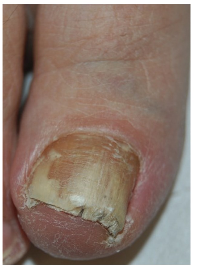
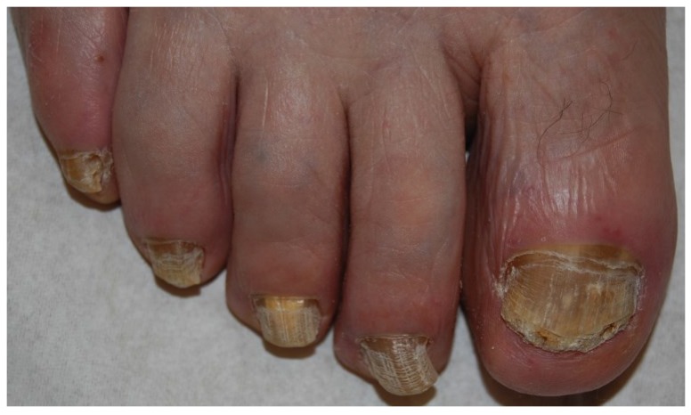
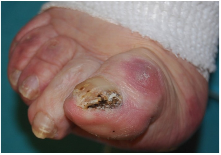
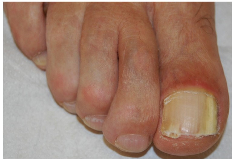
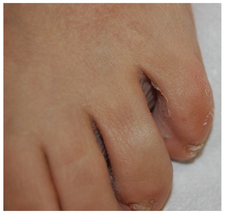
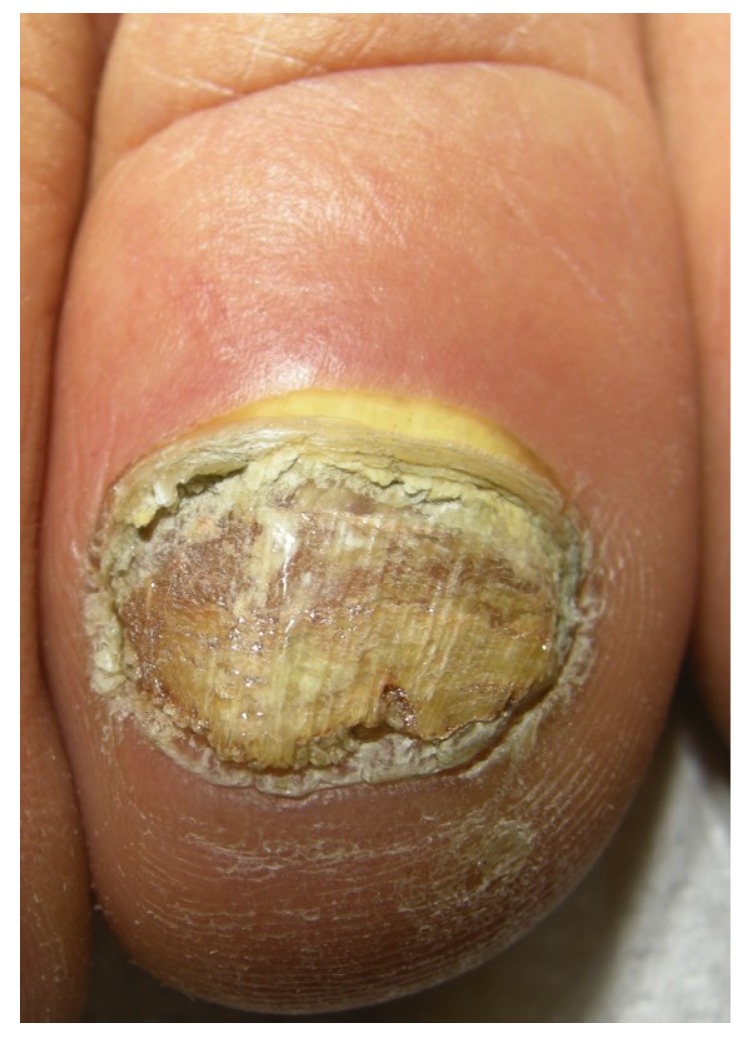
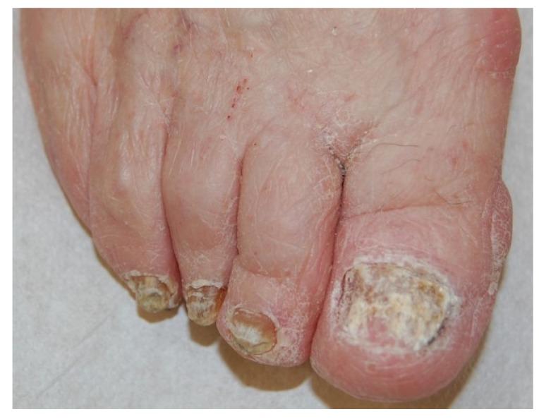
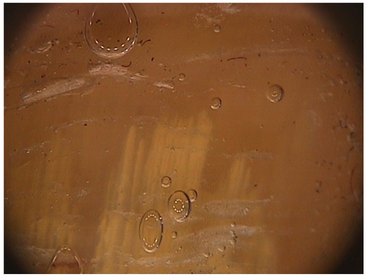
Onychomycosis is the most common nail infective disorder. It is caused mainly by anthropophilic dermatophytes, in particular by Trichophyton rubrum and T. mentagrophytes var. interdigitale. Yeasts, like Candida albicans and C. parapsilosis, and molds, like Aspergillus spp., represent the second cause of onychomycosis. The clinical suspect of onychomycosis should be confirmed my mycology. Onychoscopy is a new method that can help the physician, as in onychomycosis, it shows a typical fringed proximal margin. Treatment is chosen depending on the modality of nail invasion, fungus species and the number of affected nails. Oral treatments are often limited by drug interactions, while topical antifungal lacquers have less efficacy. A combination of both oral and systemic treatment is often the best choice.

Keywords: fungi; nail; nail lacquers; onychomycosis; systemic antifungal therapy.


Conflict of interest statement

The authors declare no conflict of interest.

Figures

Figure 1
Figure 1
Distal and lateral subungual onychomycosis (DLSO): whitish discoloration, onycholysis and subungual hyperkeratosis.
Figure 2
Figure 2
DLSO with prevalent yellow discoloration.
Figure 3
Figure 3
Pigmented DLSO.
Figure 4
Figure 4
Onychomycosis due to molds, presenting the typical periungual inflammation.
Figure 5
Figure 5
White superficial onychomycosis (WSO): white opaque friable patches of the nail plate.
Figure 6
Figure 6
Tinea pedis interdigitalis, often associated with WSO.
Figure 7
Figure 7
Proximal subungual onychomycosis (PSO): white discoloration of the proximal nail plate.
Figure 8
Figure 8
Endonyx onychomycosis: white discoloration of the nail plate that is firmly attached to the nail bed.
Figure 9
Figure 9
Total onychomycosis: the nail plate is completely invaded by fungi and friable.
Figure 10
Figure 10
Onychoscopy of DLSO, showing the typical proximal fringed (ragged) margin.

References

    1. Heikkla H., Stubb S. The prevalence of onychomycosis in Finland. Br. J. Dermatol. 1995;133:699–703. doi: 10.1111/j.1365-2133.1995.tb02741.x. - DOI - PubMed
    1. Roseeuw D. Achilles foot screening project: Preliminary results of patients screened by dermatologists. J. Eur. Acad. Dermatol. Venereol. 1999;12:S6–S9. doi: 10.1016/S0926-9959(99)00053-7. - DOI - PubMed
    1. Scher R.K., Rich P., Pariser D., Elewski B. The epidemiology, etiology, and pathophysiology of onychomycosis. Semin. Cutan. Med. Surg. 2013;32:S2–S4. - PubMed
    1. Nouripour-Sisakht S., Mirhendi H., Shidfar M.R., Ahmadi B., Rezaei-Matehkolaei A., Geramishoar M., Zarei F., Jalalizand N. Aspergillus species as emerging causative agents of onychomycosis. J. Mycol. Med. 2015 doi: 10.1016/j.mycmed.2014.12.001. - DOI - PubMed
    1. Gupta A.K., Drummond-Main C., Cooper E.A., Brintnell W., Piraccini B.M., Tosti A. Systematic review of nondermatophyte mold onychomycosis: Diagnosis, clinical types, epidemiology, and treatment. J. Am. Acad. Dermatol. 2012;66:494–502. doi: 10.1016/j.jaad.2011.02.038. - DOI - PubMed