Review
. 2018 Mar;14(3):168-181.
doi: 10.1038/nrneurol.2017.185. Epub 2018 Jan 29.

Brain insulin resistance in type 2 diabetes and Alzheimer disease: concepts and conundrums

Affiliations
Review

Brain insulin resistance in type 2 diabetes and Alzheimer disease: concepts and conundrums

Steven E Arnold et al. Nat Rev Neurol. 2018 Mar.

Abstract

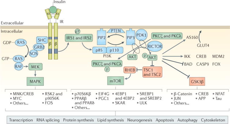
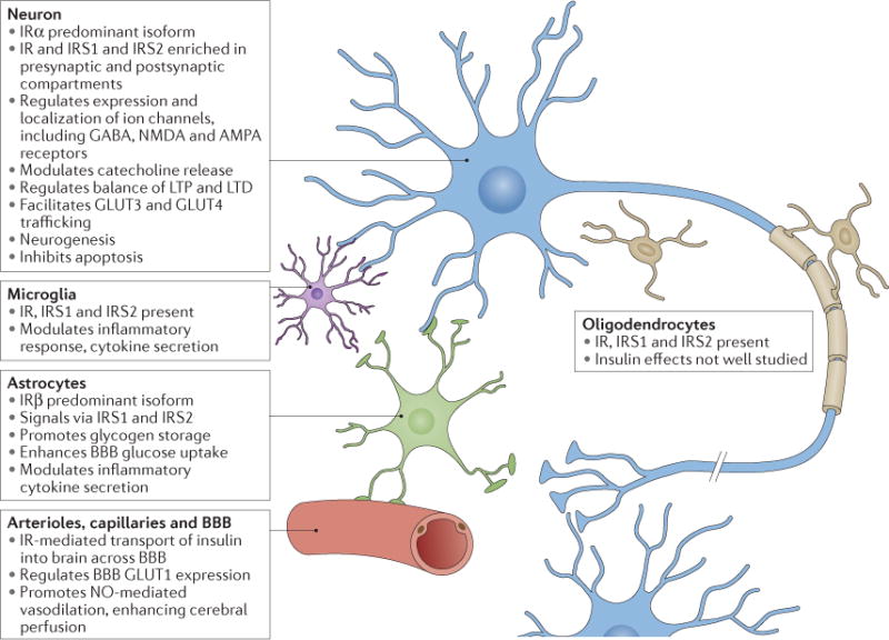
Considerable overlap has been identified in the risk factors, comorbidities and putative pathophysiological mechanisms of Alzheimer disease and related dementias (ADRDs) and type 2 diabetes mellitus (T2DM), two of the most pressing epidemics of our time. Much is known about the biology of each condition, but whether T2DM and ADRDs are parallel phenomena arising from coincidental roots in ageing or synergistic diseases linked by vicious pathophysiological cycles remains unclear. Insulin resistance is a core feature of T2DM and is emerging as a potentially important feature of ADRDs. Here, we review key observations and experimental data on insulin signalling in the brain, highlighting its actions in neurons and glia. In addition, we define the concept of 'brain insulin resistance' and review the growing, although still inconsistent, literature concerning cognitive impairment and neuropathological abnormalities in T2DM, obesity and insulin resistance. Lastly, we review evidence of intrinsic brain insulin resistance in ADRDs. By expanding our understanding of the overlapping mechanisms of these conditions, we hope to accelerate the rational development of preventive, disease-modifying and symptomatic treatments for cognitive dysfunction in T2DM and ADRDs alike.


Conflict of interest statement

Competing interests statement

The authors declare no competing interests.

Figures

Figure 1
Figure 1. Canonical insulin signalling pathways
Insulin binds extracellular α-subunits of the insulin receptor (IR), leading to dimerization and autophosphorylation of β-subunits and activation of its kinase activity. The IR phosphorylates select tyrosine residues (pY) on insulin receptor substrate 1 (IRS1) and IRS2, leading to exposure of binding sites for signalling partners. IRS1 and IRS2 recruit and activate the phosphoinositide 3-kinase (PI3K) complex, which then phosphorylates and activates AKT, the major node of the insulin signalling cascade, as well as protein kinase Cζ (PKCζ) and PKCλ. Activated AKT has many downstream effects: of greatest relevance to systemic glucose control, AKT phosphorylates AKT substrate of 160 kDa (AS160; also known as TBC1D4), which controls the translocation of glucose transporter type 4 (GLUT4) to the cell membrane for uptake of glucose into muscle, adipose and some neurons. AKT-mediated activation of mTOR and the downstream targets of mTOR serves to regulate protein and lipid synthesis and many aspects of cell metabolism, growth, survival and autophagy. Phosphorylation of glycogen synthase kinase 3β (GSK3β) by AKT inhibits the constitutive activity of this key kinase. GSK3β has many protein substrates, such as glycogen synthase, β-catenin, microtubule-associated proteins (including tau), intermediate filaments, cAMP-responsive element-binding protein (CREB) and others. Through these diverse proteins, insulin and GSK3β signalling play important parts in the regulation of cellular proliferation, migration, glucose regulation, apoptosis and neuroplasticity. AKT kinase activity also directly activates proteins such as inhibitor of nuclear factor-κB kinase (IKK), CREB and E3 ubiquitin-protein ligase Mdm2 (MDM2) to regulate transcription, cytokine production and cell survival, and it directly inhibits selected proteins, including regulators of apoptosis (Bcl2-associated agonist of cell death (BAD) and caspase 9 (CASP9)) and Forkhead box protein (FOX) transcription factors. Independent of IRS1 and IRS2 and AKT, IR kinase activity initiates the activation of the mitogen-activated protein kinase (MAPK) pathway, which is especially important for regulating the transcription of CREB, Myc proto-oncogene protein (MYC) and ribosomal protein S6 kinase 2 (RSK2; also known as S6Kα3), affecting cell proliferation, differentiation, innate and adaptive immune function and neuroplasticity. Importantly, AKT, GSK3β, mTOR and MAPK themselves provide feedback autoregulation of IRS1 and IRS2, inhibiting their activity through site-specific serine phosphorylation. 4EBP, eukaryotic translation initiation factor 4E binding protein; APP, amyloid precursor protein; EIF4G, eukaryotic translation initiation factor 4γ; FOS, proto-oncogene c-Fos; GRB2, growth factor receptor-bound protein 2; JUN, transcription factor AP-1; MEK, MAPK/ERK kinase (also known as MAPKK); MNK, MAP kinase signal-interacting kinase (also known as MKNK); NFAT, nuclear factor of activated T cells; p70S6Kβ, p70 ribosomal S6 kinase β (also known as S6Kβ2); p90S6K, 90 kDa ribosomal protein S6 kinase 1 (also known as S6Kα1); PDK1, 3-phophoinositide-dependent protein kinase 1; PGC1, PPARγ coactivator 1; PIP2, phosphatidylinositol 4,5-bisphosphate; PIP3, phosphatidylinositol (3,4,5)-trisphosphate; PPAR, peroxisome proliferator-activated receptor; RICTOR, rapamycin-insensitive companion of mTOR; SHC, SHC-transforming protein; SKAR, S6K1 Aly/REF-like target (also known as POLDIP3); SOS, son of sevenless homologue; SREBP, sterol regulatory element-binding protein; TSC1, hamartin; TSC2, tuberin.
Figure 2
Figure 2. Insulin effects in major cell types of the brain
Main characteristics of insulin signalling in neurons, astrocytes, microglia and the vascular system. AMPA, α-amino-3-hydroxy-5-methyl-4-isoxazole propionic acid; BBB, blood–brain barrier; GLUT, glucose transporter type protein; IR, insulin receptor; IRS, insulin receptor substrate; LTD, long-term depression; LTP, long-term potentiation; NMDA, N-methyl-d-aspartate; NO, nitric oxide.

References

    1. Snyder HM, et al. Vascular contributions to cognitive impairment and dementia including Alzheimer’s disease. Alzheimers Dement. 2015;11:710–717. - PMC - PubMed
    1. Montine TJ, et al. Recommendations of the Alzheimer’s disease-related dementias conference. Neurology. 2014;83:851–860. - PMC - PubMed
    1. Stoeckel LE, et al. Complex mechanisms linking neurocognitive dysfunction to insulin resistance and other metabolic dysfunction. F1000Res. 2016;5:353. - PMC - PubMed
    1. Chatterjee S, et al. Type 2 diabetes as a risk factor for dementia in women compared with men: a pooled analysis of 2.3 million people comprising more than 100,000 cases of dementia. Diabetes Care. 2016;39:300–307. - PMC - PubMed
    1. Gao C, Liu Y, Li L, Holscher C. New animal models of Alzheimer’s disease that display insulin desensitization in the brain. Rev Neurosci. 2013;24:607–615. - PubMed

Publication types