Review
. 2018 Apr;1418(1):5-19.
doi: 10.1111/nyas.13531. Epub 2018 Jan 28.

Misfolded proinsulin in the endoplasmic reticulum during development of beta cell failure in diabetes

Affiliations
Review

Misfolded proinsulin in the endoplasmic reticulum during development of beta cell failure in diabetes

Anoop Arunagiri et al. Ann N Y Acad Sci. 2018 Apr.

Abstract

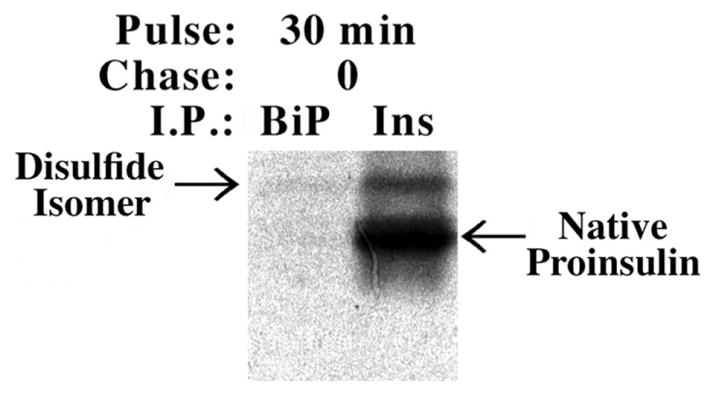
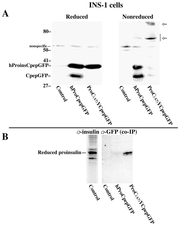
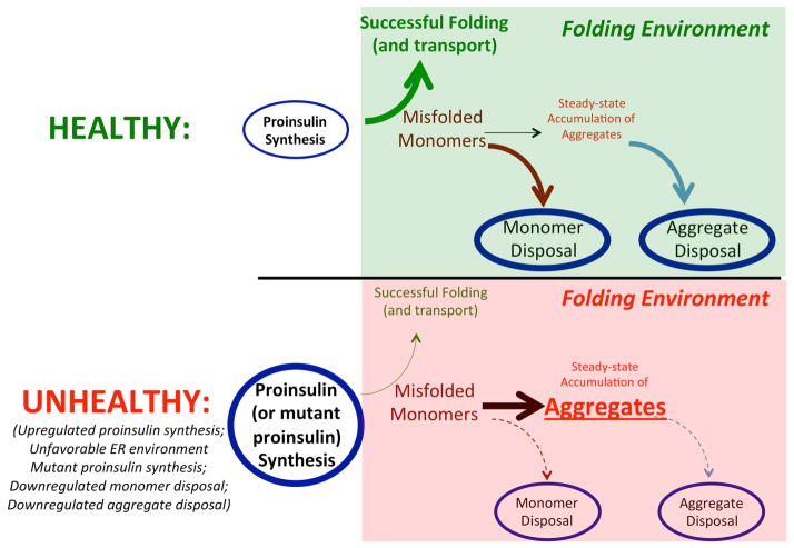
The endoplasmic reticulum (ER) is broadly distributed throughout the cytoplasm of pancreatic beta cells, and this is where all proinsulin is initially made. Healthy beta cells can synthesize 6000 proinsulin molecules per second. Ordinarily, nascent proinsulin entering the ER rapidly folds via the formation of three evolutionarily conserved disulfide bonds (B7-A7, B19-A20, and A6-A11). A modest amount of proinsulin misfolding, including both intramolecular disulfide mispairing and intermolecular disulfide-linked protein complexes, is a natural by-product of proinsulin biosynthesis, as is the case for many proteins. The steady-state level of misfolded proinsulin-a potential ER stressor-is linked to (1) production rate, (2) ER environment, (3) presence or absence of naturally occurring (mutational) defects in proinsulin, and (4) clearance of misfolded proinsulin molecules. Accumulation of misfolded proinsulin beyond a certain threshold begins to interfere with the normal intracellular transport of bystander proinsulin, leading to diminished insulin production and hyperglycemia, as well as exacerbating ER stress. This is most obvious in mutant INS gene-induced Diabetes of Youth (MIDY; an autosomal dominant disease) but also likely to occur in type 2 diabetes owing to dysregulation in proinsulin synthesis, ER folding environment, or clearance.

Keywords: ER-associated degradation; Mutant INS gene-induced Diabetes of Youth; MIDY; disulfide mispairing; endoplasmic reticulum stress; ER; protein aggregation; secretory protein synthesis.


Conflict of interest statement

Competing Interests

The authors declare that we have no competing interests related to this work.

Figures

Figure 1
Figure 1
Pattern of disulfide bond formation throughout the insulin/IGF superfamily. (A) Reproduced from Ref. , with permission from the publisher. (B) The human insulin peptide sequence is inserted into the disulfide pattern shown in panel A. The first two Cys residues fall within the insulin B-chain (in red), and the remaining four Cys residues fall within the insulin A-chain (in blue). The conversion of the single-chain proinsulin to the two-chain insulin molecule involves excision of the connecting C-peptide (shown in orange) via endoproteolytic cleavage followed by carboxypeptidase cleavage at dibasic sites (green letters).
Figure 2
Figure 2
Non-native isomer of newly synthesized proinsulin shows enhanced binding to the ER HSP70 chaperone BiP. Fifty isolated rat pancreatic islets were pulse labeled with 35S-labeled amino acids and then lysed immediately and either immunoprecipitated with a polyclonal antibody to insulin that recognizes all proinsulin forms or co-immunoprecipitated with anti-BiP. The immunoprecipitates were analyzed by nonreducing Tris–Tricine–urea–SDS-PAGE. In addition to native disulfide-bonded proinsulin, a more slowly migrating proinsulin disulfide isomer is preferentially associated with BiP. Reproduced from Ref. with permission from the publisher.
Figure 3
Figure 3
Protein interactions of proinsulin-C(A7)Y. (A) The INS1 pancreatic beta cell line was used, either untransfected (control) or transfected to express hPro-CpepGFP or hProC(A7)Y-CpepGFP (the latter bearing the Akita proinsulin mutation). Cell lysates were subjected to western blotting with anti-GFP after SDS-PAGE under reduced or nonreduced conditions. Not only is hProC(A7)Y-CpepGFP not endoproteolytically processed in beta cells, but the protein is recovered in higher-molecular-mass protein complexes (open arrows) that are detected only under nonreduced conditions. (B) The same cells from panel A were pulse labeled with 35S-labeled amino acids for 30 min and lysed, then GFP-containing peptides were immunoprecipitated, and the samples were analyzed by Tris–tricine–urea–SDS-PAGE under reducing conditions to detect coimmunoprecipitation of endogenous proinsulin. Reproduced from Ref. (© 2007; National Academy of Sciences).
Figure 4
Figure 4
Steps contributing to proinsulin aggregation in the ER folding environment. Upper panel: under healthy conditions, proinsulin synthesis is limited to physiological levels (small font) and folds within a generally favorable folding environment leading to successful export from the ER (thick green arrow). The misfolded proinsulin that is generated in parallel with native folding is actively disposed of, including monomer disposal (thick brown arrow) and aggregate disposal (thick blue arrow). Through each of these mechanisms, the steady-state level of misfolded proinsulin is held to low levels. Lower panel: under unhealthy conditions, proinsulin synthesis is exuberant to a level that may be considered supraphysiological; the increased supply of unfolded monomers leads to the production of more proinsulin aggregates, exceeding the disposal of misfolded proinsulin, such that the steady-state level of misfolded proinsulin is increased.
Figure 5
Figure 5
Glucose-related changes in eIF2 phosphorylation in MIN6 cells. MIN6 cells (i.e., cells of a mouse pancreatic beta cell line) were preincubated in no glucose (1 h) before incubation in the indicated concentrations of glucose for 1 h in the presence of [35S]methionine. Upper panel: cell lysates were analyzed by SDS-PAGE and western blotting with anti-phospho-eIF2α (P-eIF2α) and anti-eIF2α. Lower panel: incorporation of [35S]methionine into total protein, as a percent of control (0 mm glucose). Adapted from Ref. with permission from the publisher.
Figure 6
Figure 6
Rapid formation of native proinsulin disulfide bonds. Isolated mouse pancreatic islets were pulse labeled for 60 s with 35S-labeled amino acids and either lysed without chase or chased in the presence of cycloheximide (100 μg/mL) to prevent further proinsulin synthesis. The islets were lysed and analyzed by Tris–-tricine–urea–SDS-PAGE and phosphorimaging; the position of oxidized native proinsulin is shown. Reproduced from Ref. with permission from the publisher.
Figure 7
Figure 7
Hypothesis: relevance of proinsulin misfolding to insulin deficiency/beta cell dysfunction in garden-variety diabetes mellitus. This figure has been adapted and altered from Fig. 1 of Ref. ; permission from publisher is pending.

References

    1. Laybutt DR, Preston AM, Akerfeldt MC, et al. Endoplasmic reticulum stress contributes to beta cell apoptosis in type 2 diabetes. Diabetologia. 2007;50:752–763. - PubMed
    1. Fonseca SG, Gromada J, Urano F. Endoplasmic reticulum stress and pancreatic beta-cell death. Trends Endocrinol Metab. 2011;22:266–274. - PMC - PubMed
    1. Eizirik DL, Cnop M. ER stress in pancreatic beta cells: the thin red line between adaptation and failure. Sci Signal. 2010;3:pe7. - PubMed
    1. Oakes SA, Papa FR. The role of endoplasmic reticulum stress in human pathology. Annu Rev Pathol. 2015;10:173–194. - PMC - PubMed
    1. Wang M, Kaufman RJ. Protein misfolding in the endoplasmic reticulum as a conduit to human disease. Nature. 2016;529:326–335. - PubMed

Publication types