

Human placental oxygenation in late gestation: experimental and theoretical approaches
- PMID: 29377190
- PMCID: PMC6265570
- DOI: 10.1113/JP275633
Human placental oxygenation in late gestation: experimental and theoretical approaches
Abstract
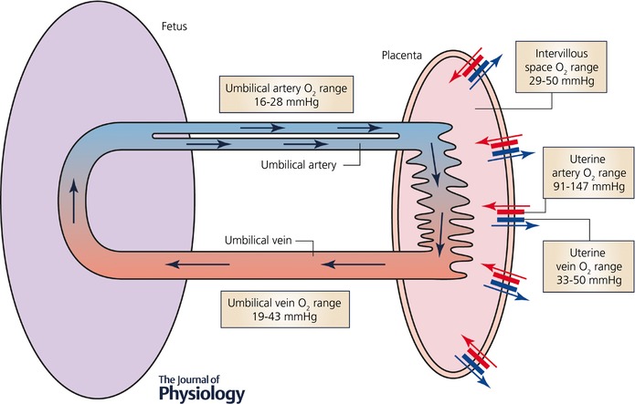
The placenta is crucial for life. It is an ephemeral but complex organ acting as the barrier interface between maternal and fetal circulations, providing exchange of gases, nutrients, hormones, waste products and immunoglobulins. Many gaps exist in our understanding of the detailed placental structure and function, particularly in relation to oxygen handling and transfer in healthy and pathological states in utero. Measurements to understand oxygen transfer in vivo in the human are limited, with no general agreement on the most appropriate methods. An invasive method for measuring partial pressure of oxygen in the intervillous space through needle electrode insertion at the time of Caesarean sections has been reported. This allows for direct measurements in vivo whilst maintaining near normal placental conditions; however, there are practical and ethical implications in using this method for determination of placental oxygenation. Furthermore, oxygen levels are likely to be highly heterogeneous within the placenta. Emerging non-invasive techniques, such as MRI, and ex vivo research are capable of enhancing and improving current imaging methodology for placental villous structure and increase the precision of oxygen measurement within placental compartments. These techniques, in combination with mathematical modelling, have stimulated novel cross-disciplinary approaches that could advance our understanding of placental oxygenation and its metabolism in normal and pathological pregnancies, improving clinical treatment options and ultimately outcomes for the patient.
Keywords: FGR; MRI; intervillious space; modelling; oxygen; perfusion; placenta.
© 2018 University of Oxford. The Journal of Physiology published by John Wiley & Sons Ltd on behalf of The Physiological Society.
Figures
References
-
- Arikan GM, Scholz HS, Petru E, Haeusler MC, Haas J & Weiss PA (2000). Cord blood oxygen saturation in vigorous infants at birth: what is normal? BJOG 107, 987–994. - PubMed
-
- Bernardez‐Zapata FJ & Moreno‐Rey C (2014). [Normal values of gases in the vein of the umbilical cord during the postpartum period and postcesarea immediately in normal fetuses to term]. Ginecol Obstet Mex 82, 170–176. - PubMed
-
- Blechner JN, Cotter JR, Stenger VG, Hinkley CM & Prystowsky H (1968). Oxygen, carbon dioxide, and hydrogen ion concentrations in arterial blood during pregnancy. Am J Obstet Gynecol 100, 1–6. - PubMed
-
- Brownbill P, Sebire N, McGillick EV, Ellery S & Murthi P (2018). Ex vivo dual perfusion of the human placenta: disease simulation, therapeutic pharmacokinetics and analysis of off‐target effects. Methods Mol Biol 1710, 173–189. - PubMed
Publication types
MeSH terms
Substances
Grants and funding
LinkOut - more resources
Full Text Sources
Other Literature Sources
Miscellaneous