. 2018 Jan 29;14(1):e1007183.
doi: 10.1371/journal.pgen.1007183. eCollection 2018 Jan.

Rewired RNAi-mediated genome surveillance in house dust mites

Affiliations

Rewired RNAi-mediated genome surveillance in house dust mites

Mosharrof Mondal et al. PLoS Genet. .

Abstract

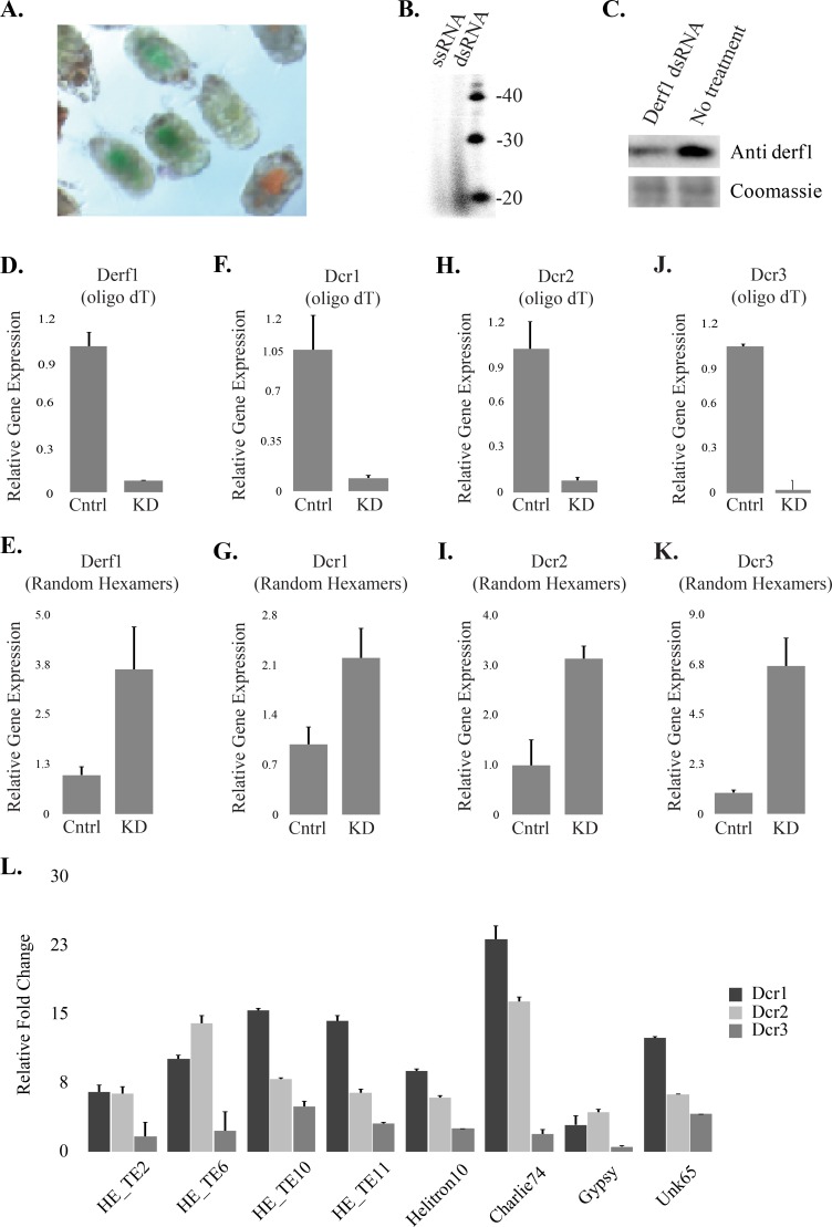
House dust mites are common pests with an unusual evolutionary history, being descendants of a parasitic ancestor. Transition to parasitism is frequently accompanied by genome rearrangements, possibly to accommodate the genetic change needed to access new ecology. Transposable element (TE) activity is a source of genomic instability that can trigger large-scale genomic alterations. Eukaryotes have multiple transposon control mechanisms, one of which is RNA interference (RNAi). Investigation of the dust mite genome failed to identify a major RNAi pathway: the Piwi-associated RNA (piRNA) pathway, which has been replaced by a novel small-interfering RNA (siRNA)-like pathway. Co-opting of piRNA function by dust mite siRNAs is extensive, including establishment of TE control master loci that produce siRNAs. Interestingly, other members of the Acari have piRNAs indicating loss of this mechanism in dust mites is a recent event. Flux of RNAi-mediated control of TEs highlights the unusual arc of dust mite evolution.


Conflict of interest statement

The authors have declared that no competing interests exist.

Figures

Fig 1
Fig 1. Absence of Piwi/piRNA pathway in dust mites.
A. Relationship of Ago/Piwi proteins from D. farinae, Drosophila, C. elegans, and A. suum using conserved Paz, Mid and Piwi domains. Dust mite proteins indicated in red. Only two Wago proteins included for simplicity. Bootstrap values for major nodes indicated. B-D. Heatmaps showing Z-scores for Overlap probabilities for 18-30nt small RNAs from dust mites (B), spider mites (C), and Drosophila female bodies (D). Overlaps are shown for each read length as well as all lengths together. Read lengths listed horizontally, Overlaps vertically. Blue arrow labeled Dcr indicates expected 2nt register suggestive of dicer cleavage. Red arrow labeled pp shows expected overlap for ping-pong processing.
Fig 2
Fig 2. Distribution of small RNA mapping across dust mite genomic features.
A. An RDI plot of per locus strand bias seen after multi-mapping and uniquely-mapping protocols in dust mite genome feature classes: mRNA, rRNA, TE, tRNA, U6 snRNA, and unknown genomic loci. Mean indicated by black bar, white transparent box shows standard deviation. Under the graph millions of reads and number of loci in each category is shown. B. Coverage of small RNA mapping in TEs (left) and mRNAs (right). Line plots show average coverage across loci. Heatmaps below show length-normalized per locus coverage of small RNA reads.
Fig 3
Fig 3. siRNAs facilitate genome surveillance in dust mite.
A. Size distribution of genome mapped small RNAs in dust mites (solid line) and spider mites (dashed line). B. Size distribution of TE mapped RNAs small in dust mites (solid line) and spider mites (dashed line). C. Seqlogo showing 5’ nucleotide bias in TE mapped small RNA in spider mites (top) and dust mites (bottom). D. Per locus biogenesis of dust mite TE associated small RNAs. Left shows Log2 read accumulation per read size. Overhang probabilities (positive z-scores only) of small RNA pairs at specific or all sizes. The size(s) of reads show above heat map. A similar analysis from spider mite small RNAs (18-30nt) shown on right. Red arrow indicates overlap for ping pong process. Blue arrow shows overlap expected for dicer processing.
Fig 4
Fig 4. RNAi in dust mites.
A. Dust mite soaking. Mites were soaked separately in orange and green food color for 30 min. B. Radiolabeled RNAs recovered from mites fed either single-stranded (ssRNA) or double-stranded RNA (dsRNA). RNAs were treated with DNase and CIP prior to separation via denaturing PAGE. C. Western blot of Derf1 allergen after soaking animals with derf1 dsRNA (upper panel) and coomassie staining of the membrane (lower panel). Animals were soaked for 30 min and after 4 days lysates were prepared. D-K. qPCR for dust mite transcripts, all experiments were performed at least three times. Values represent four technical replicates. Reverse transcription was carried out with either oligo dT (D, F, H, J) or with random hexamers (E, G, I, K). Target transcripts were derf1 (D,E), dcr1 (F,G), dcr2 (H, I), and dcr3 (J, K). Cntrl represents no treatment, and KD soaking in the indicated dsRNA. L. Increased expression of numerous TE’s (S2 Table) following RNAi against three dust mite Dicers relative to untreated control. Error bars represent SEM.
Fig 5
Fig 5. Characteristics of small RNA-mediated TE control in dust mites.
A. siRNA producing TE-control master loci (ML). Read density of all mapping events to the positive strand in red, negative strand in blue. Density of uniquely mapping reads in yellow for positive strand and green for negative strand. B. Catalog of TE homology sequences in master loci. Multiple sequence alignment of TEs against master loci to show homologous sequences. C. Northern blots against ML-associated siRNAs (ML-A siRNA) after eliciting RNAi against dust mite Dicers. D. Northern blots against ML-A siRNAs after β-elimination test. E. Accumulation of ML-A siRNAs following incubation with the monophosphate specific terminator ribonuclease (term) and Calf intestinal phosphatase (CIP). Relative accumulation of ML-A siRNAs was determined by densitometry and normalization to U6 signal. Experiments were performed at least three times, representative results shown.
Fig 6
Fig 6. DNA methylation status in dust mite.
A. Distribution of methylated bases assessed by bisulfite sequencing across the entire genome, mRNAs, and TEs. Percentage of methylated Cs (mC) identified in all sequence contexts are compared with the number of bases identified in each category. B. Dust Mite DNMT1 homolog. Expression of dust mite DNA (cytosine-5)-methyltransferase 1 (Dnmt1) in mixed stage RNA-Seq data. Blue bar represents dust mite Dnmt1 locus in the scaffold. Read density in region shown as grey plot. Reads mapping below; plus strand mapping in red, minus strand mapping in blue. C. Domain structure of truncated D. farinae Dnmt1 and an intact ortholog from Limulus polyphemus.

Comment in

References

    1. Arlian LG. House-dust-mite allergens: a review. Exp Appl Acarol. 1991;10(3–4):167–86. . - PubMed
    1. Klimov PB, OC B. Is permanent parasitism reversible?—critical evidence from early evolution of house dust mites. Syst Biol. 2013;62(3):411–23. doi: 10.1093/sysbio/syt008 . - DOI - PubMed
    1. Brookfield JF. Host-parasite relationships in the genome. BMC Biol. 2011;9:67 doi: 10.1186/1741-7007-9-67 ; PubMed Central PMCID: PMCPMC3189907. - DOI - PMC - PubMed
    1. Poulin R, Randhawa HS. Evolution of parasitism along convergent lines: from ecology to genomics. Parasitology. 2015;142 Suppl 1:S6–S15. doi: 10.1017/S0031182013001674 ; PubMed Central PMCID: PMCPMC4413784. - DOI - PMC - PubMed
    1. Fedoroff NV. Presidential address. Transposable elements, epigenetics, and genome evolution. Science. 2012;338(6108):758–67. . - PubMed

Publication types

Substances