. 2018 Jan 29;13(1):e0191653.
doi: 10.1371/journal.pone.0191653. eCollection 2018.

16S rRNA gene metabarcoding and TEM reveals different ecological strategies within the genus Neogloboquadrina (planktonic foraminifer)

Affiliations

16S rRNA gene metabarcoding and TEM reveals different ecological strategies within the genus Neogloboquadrina (planktonic foraminifer)

Clare Bird et al. PLoS One. .

Abstract

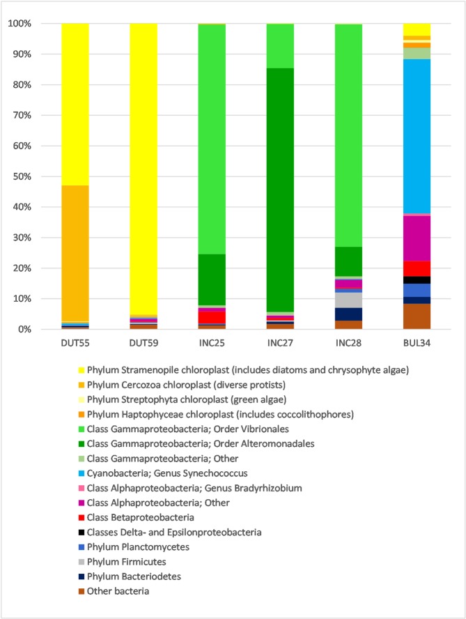
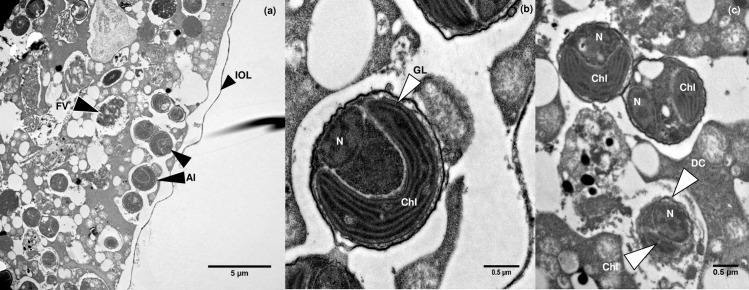
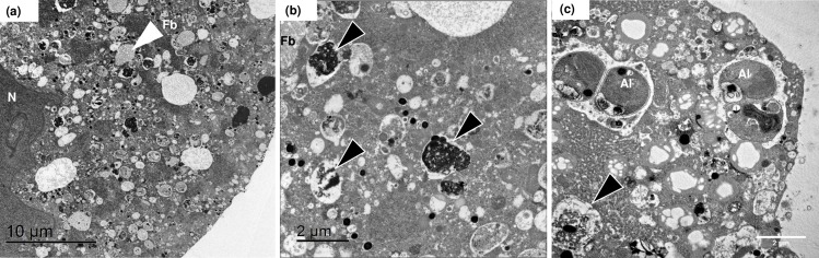
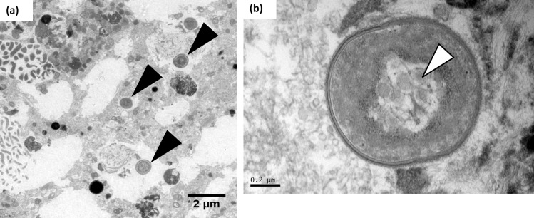
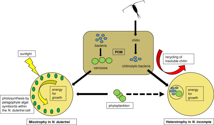
Uncovering the complexities of trophic and metabolic interactions among microorganisms is essential for the understanding of marine biogeochemical cycling and modelling climate-driven ecosystem shifts. High-throughput DNA sequencing methods provide valuable tools for examining these complex interactions, although this remains challenging, as many microorganisms are difficult to isolate, identify and culture. We use two species of planktonic foraminifera from the climatically susceptible, palaeoceanographically important genus Neogloboquadrina, as ideal test microorganisms for the application of 16S rRNA gene metabarcoding. Neogloboquadrina dutertrei and Neogloboquadrina incompta were collected from the California Current and subjected to either 16S rRNA gene metabarcoding, fluorescence microscopy, or transmission electron microscopy (TEM) to investigate their species-specific trophic interactions and potential symbiotic associations. 53-99% of 16S rRNA gene sequences recovered from two specimens of N. dutertrei were assigned to a single operational taxonomic unit (OTU) from a chloroplast of the phylum Stramenopile. TEM observations confirmed the presence of numerous intact coccoid algae within the host cell, consistent with algal symbionts. Based on sequence data and observed ultrastructure, we taxonomically assign the putative algal symbionts to Pelagophyceae and not Chrysophyceae, as previously reported in this species. In addition, our data shows that N. dutertrei feeds on protists within particulate organic matter (POM), but not on bacteria as a major food source. In total contrast, of OTUs recovered from three N. incompta specimens, 83-95% were assigned to bacterial classes Alteromonadales and Vibrionales of the order Gammaproteobacteria. TEM demonstrates that these bacteria are a food source, not putative symbionts. Contrary to the current view that non-spinose foraminifera are predominantly herbivorous, neither N. dutertrei nor N. incompta contained significant numbers of phytoplankton OTUs. We present an alternative view of their trophic interactions and discuss these results within the context of modelling global planktonic foraminiferal abundances in response to high-latitude climate change.


Conflict of interest statement

Competing Interests: The authors have declared that no competing interests exist.

Figures

Fig 1
Fig 1. Relative abundance of taxonomically assigned 16S rRNA gene sequences from six individual foraminifer specimens.
Two N. dutertrei (DUT55 and DUT59), three N. incompta specimens (INC25, INC27 and INC28) and one G. bulloides specimen (BUL34; [59]) are shown for comparison. Sequences are assigned to operational taxonomic units (OTUs) and have been grouped in the figure at higher levels of taxonomic classification for visual clarity (see key). Assignation of lower taxonomic ranks are discussed in the text, such as the OTUs grouped within the Gammaproteobacteria, in the order Vibrionales.
Fig 2
Fig 2. Fluorescence micrograph of a DAPI-stained decalcified N. dutertrei cell.
(a) Diffuse autofluorescence can be observed throughout the cytoplasm. The observed ~2 μm diameter structures prevalent throughout the cell are consistent with the presence of picoeukaryotic algae. The white arrowhead denotes an example of the bright spots, 5–10μm in size that may be food vacuoles containing condensed prey items. The white rectangle denotes the area magnified in (b) where the black arrowhead highlights one of the ~2 μm diameter putative picoeukaryotes.
Fig 3
Fig 3. Fluorescence micrographs of decalcified N. incompta and G. bulloides cells.
(a) High levels of background autofluorescence can be observed throughout the cytoplasm of the DAPI-stained decalcified N. incompta cell. However, in contrast to N. dutertrei (Fig 2), there are no abundant algal nuclei observable above background signals. In addition, in contrast to decalcified G. bulloides, where cyanobacterial cells can easily be detected (b) no bacterial cells can be observed in N. incompta.
Fig 4
Fig 4. Transmission electron micrographs of pelagophyte cells inside N. dutertrei.
(a) Intact picoeukaryotic algae can be observed (Al) in close proximity to the cell periphery. The inner organic layer (IOL) is also clearly visible. A probable food vacuole can also be seen (Fv). (b) An individual algal cell with a clearly visible horse-shoe shaped chloroplast (Chl) with girdle lamella (GL) and discernible nucleus (N). (c) Two intact algal cells with obvious chloroplasts and nuclei can be seen above an algal cell being digested (DC). The nucleus and chloroplast of this cell are still observable, but the cell membrane appears no longer to be fully intact.
Fig 5
Fig 5. Transmission electron micrographs of N. incompta.
(a) Low-magnification region of N. incompta showing the nucleus (N) and a fibrillar body (fb). (b) Higher magnification image shows a fibrillar body (Fb) and probable food vacuoles (black arrowheads). (c) A very small number of algal cells in early degradation (Al) were observed in some N. incompta individuals.
Fig 6
Fig 6. Transmission electron micrographs of G. bulloides.
(a) The black arrowheads depict cyanobacteria of the genus Synechococcus [59] within the G. bulloides cytoplasm. (b) Higher magnification of a Synechococcus endobiont, with visible characteristic carboxysomes (white arrowhead).
Fig 7
Fig 7. Proposed trophic interactions of N. dutertrei and N. incompta.
The illustrated trophic interactions are based on 16S rRNA gene metabarcoding performed in this study and laboratory observations reported in the literature. Black arrows indicate direction of heterotrophic carbon flow. We propose that N. dutertrei Type Ic is a mixotrophic feeder. Pelagophyte algae provide energy for growth through photosynthesis, either via direct consumption of the algae and/or by cross-membrane transport of photosynthates. In addition, N. dutertrei feeds on other protists and phytoplankton, either from the water column or from inside the POM feeding cyst. For example, within the feeding cyst N. dutertrei consumes Cercozoa that graze on the POM associated bacteria. In contrast, we propose that N. incompta Type II is a heterotrophic feeder. As zooplankton moult their chitin carapaces or die, their chitin becomes incorporated into the POM. Here chitinoclastic bacteria (eg. orders Vibrionales and Alteromonadales) break down the chitin to utilise the C and N source. N. incompta feeds on these orders of bacteria within its POM feeding cyst. N. incompta also feeds minimally on phytoplankton, but whether from the water column and/or from the feeding cyst is unknown (dashed black arrow).

References

    1. Leal M, Ferrier-Pagès C. Molecular trophic markers in marine food webs and their potential use for coral ecology. Mar Genomics. 2016; 29:1–7. doi: 10.1016/j.margen.2016.02.003 - DOI - PubMed
    1. Pompanon F, Deagle BE, Symondson WOC, Brown DS, Jarman SN, Taberlet P. Who is eating what: diet assessment using next generation sequencing. Mol Ecol. 2012; 21:1931–1950. doi: 10.1111/j.1365-294X.2011.05403.x - DOI - PubMed
    1. Ray JL, Althammer J, Skaar KS, Simonelli P, Larsen A, Stoecker D, et al. Metabarcoding and metabolome analyses of copepod grazing reveal feeding preference and linkage to metabolite classes in dynamic microbial plankton communities. Mol Ecol. 2016; 25:5585–5602. doi: 10.1111/mec.13844 - DOI - PubMed
    1. Zhang H, Xu Q, Zhao Y, Yang H. Sea cucumber (Apostichopus japonicus) eukaryotic food source composition determined by 18s rDNA barcoding. Mar Biol. 2016; 163:153 doi: 10.1007/s00227-016-2931-x - DOI
    1. Azam F, Smith DC, Steward GF, Hagstrӧm Å. Bacteria-organic matter coupling and its significance for oceanic carbon cycling. Microb Ecol. 1994; 28:167–179. doi: 10.1007/BF00166806 - DOI - PubMed

Publication types

MeSH terms

Substances

LinkOut - more resources