Review
. 2018 Feb 27;12(2):912-931.
doi: 10.1021/acsnano.7b05876. Epub 2018 Feb 6.

Nanoparticles for Immune Cytokine TRAIL-Based Cancer Therapy

Affiliations
Review

Nanoparticles for Immune Cytokine TRAIL-Based Cancer Therapy

Pedro P G Guimarães et al. ACS Nano. .

Abstract

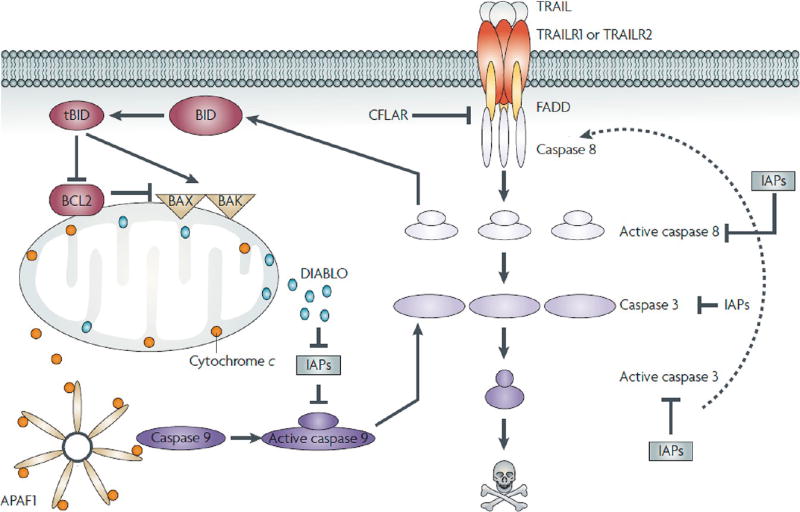
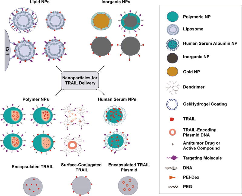
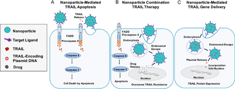
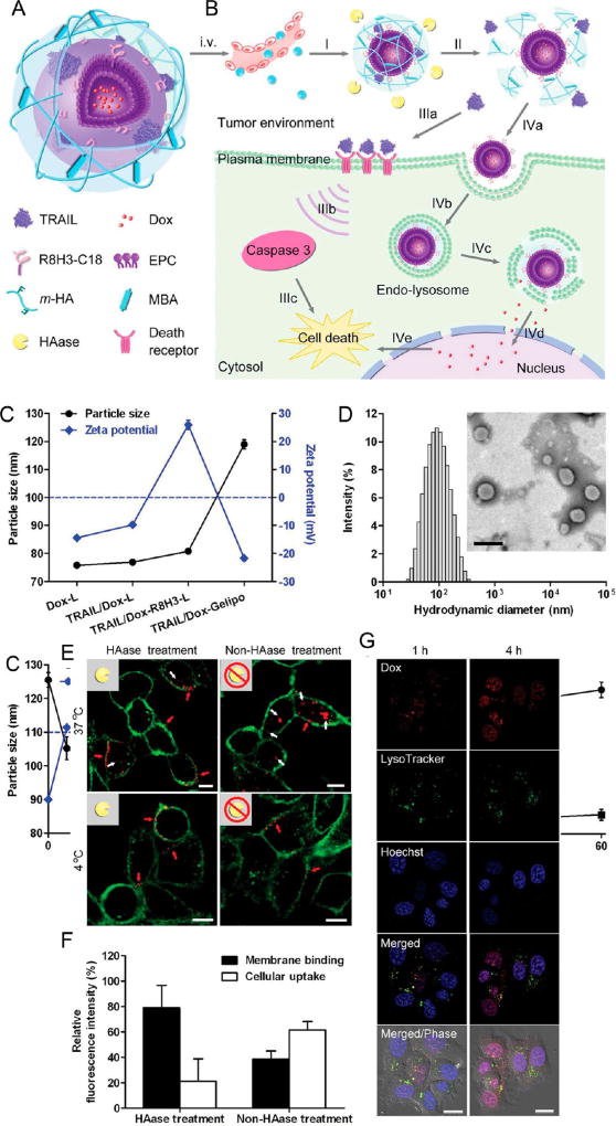
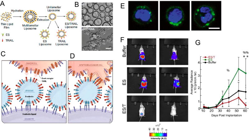
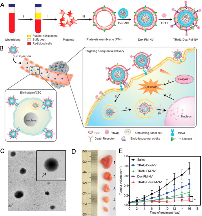
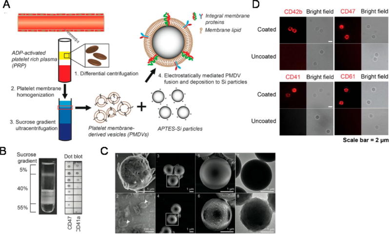
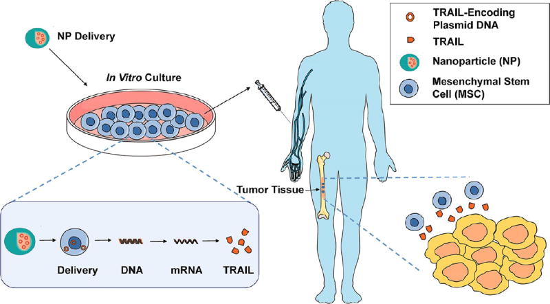
The immune cytokine tumor necrosis factor-related apoptosis-inducing ligand (TRAIL) has received significant attention as a cancer therapeutic due to its ability to selectively trigger cancer cell apoptosis without causing toxicity in vivo. While TRAIL has demonstrated significant promise in preclinical studies in mice as a cancer therapeutic, challenges including poor circulation half-life, inefficient delivery to target sites, and TRAIL resistance have hindered clinical translation. Recent advances in drug delivery, materials science, and nanotechnology are now being exploited to develop next-generation nanoparticle platforms to overcome barriers to TRAIL therapeutic delivery. Here, we review the design and implementation of nanoparticles to enhance TRAIL-based cancer therapy. The platforms we discuss are diverse in their approaches to the delivery problem and provide valuable insight into guiding the design of future nanoparticle-based TRAIL cancer therapeutics to potentially enable future translation into the clinic.

Keywords: biological barriers; biomaterials; cancer therapy; drug delivery; gene therapy; immunotherapy; metastasis; nanotechnology; oncology; tumor targeting.


Conflict of interest statement

The authors declare no competing financial interest.

Figures

Figure 1
Figure 1
Apoptotic signaling through the tumor necrosis factor (TNF)-related apoptosis-inducing ligand (TRAIL) pathway. Binding of TRAIL to TRAIL receptor 1 (TRAILR1 or D4) or TRAILR2/DR5 results in receptor oligomerization and recruitment of FAS-associated protein with death domain (FADD) and caspase 8 to form a functional death-inducing signaling complex (DISC). Upon DISC formation, caspase 8 is cleaved and activated, which in turn can cleave and activate caspase 3 and the BH3-only protein BID. Active, cleaved BID (tBID) can bind to pro-apoptotic Bax and Bak, resulting in mitochondrial membrane permeabilization and release of mitochondrial proteins cytochrome c and DIABLO. Cytochrome c, apoptotic protease-activating factor 1 (APAF1), and caspase 9 combine with ATP to form a functional apoptosome that results in cleavage and activation of caspase 9, which can then cleave caspase 3. DIABLO suppresses the caspase-inhibitory activities of inhibitor of apoptosis proteins. Caspase 3 can cleave a large number of intracellular targets, resulting in the morphological and biochemical hallmarks of apoptosis. Caspase 3 can also cleave and activate caspase 8, thereby amplifying the apoptotic signal. Reprinted with permission from ref . Copyright 2016 Nature.
Figure 2
Figure 2
Examples of classes of NPs used for TRAIL-based cancer therapy.
Figure 3
Figure 3
Approaches to utilize NPs to induce TRAIL-mediated apoptosis in tumor cells via (A) TRAIL-coated NPs binding to the tumor cell surface, (B) a combination NP-based approach that delivers encapsulated small molecule drugs to sensitize tumor cells to TRAIL-mediated apoptosis, or (C) gene delivery to cells to induce secretion of TRAIL.
Figure 4
Figure 4
Schematic design of TRAIL/Dox-Gelipo for sequential and site-specific drug delivery. (A) Main components of TRAIL/Dox-Gelipo: R8H3-modified liposomal core loading doxorubicin (Dox) and cross-linked hyaluronic acid (HA) gel-based outer shell encapsulating TRAIL. (B) Sequential delivery of TRAIL to the plasma membrane and Dox to the nuclei by TRAIL/Dox-Gelipo for combination cancer treatment. I, accumulation of Gelipo (blue balls) at the tumor site through the passive and active targeting effects; II, degradation of HA cross-linked shell by HAase; IIIa, released TRAIL binding onto the death receptors on the plasma membrane; IIIb, activation of the caspase 3 signaling pathway; IIIc, induction of the cell death; IVa, exposure of R8H3 facilitating the tumor cellular uptake of Dox-R8H3-L; IVb, internalization of Dox-R8H3-L into the tumor cells; IVc, endolysosomal escape; IVd, accumulation of the released Dox into nucleus; IVe, intercalation of Dox on DNA inducing the cell death. (C) Particle size and ζ-potential of Dox-L, TRAIL/Dox-L, TRAIL/Dox-R8H3-L, and TRAIL/Dox-Gelipo. (D) Hydrodynamic size of TRAIL/Dox-Gelipo measured by dynamic light scattering. Inset: TEM image of TRAIL/Dox-Gelipo. Scale bar is 200 nm. (E) Membrane binding efficiency of r-TRAIL from r-TRAIL-Gelipo with and without 30 min of HAase pretreatment on MDA-MB-231 cells observed by confocal laser scanning microscopy (CLSM). The plasma membranes were stained by Alexa Fluor 488 conjugate of wheat germ agglutinin. Scale bars are 10 µm. (F) Quantitative analysis on the r-TRAIL amount on the plasma membrane and in the cells. (G) Intracellular delivery of TRAIL/Dox-Gelipo after 30 min of HAase pretreatment on MDA-MB-231 cells at different time observed by CLSM. The late endosomes and lysosomes were stained by LysoTracker Green, and the nuclei were stained by Hoechst 33342. Scale bars are 20 µm. Reprinted with permission from ref . Copyright 2014 Wiley.
Figure 5
Figure 5
(A) Schematic illustration of PEI-Dexa synthesis and (B) nucleus-targeted sandwich-type Au-PEI/DNA/PEI-Dexa complex for intercellular DNA delivery. Transmission electron microscopic images of (C) Au-PEI, (D) Au-PEI/DNA, and (E) Au-PEI/DNA/PEI-Dexa. (Insets) Higher magnification micrographs of the assembled NPs. (F) Intracellular localization of PEI/DNA, Au-PEI/DNA/PEI (w/w/w, 0.6/1/2), and Au-PEI/DNA/PEI-Dexa (w/w/w, 0.6/1/2) in Hep3B cells after gene transfection for 2 or 6 h (red, Cy5-labeled pDNA; blue, DAPI stained cell nuclei; pink, colocalization). A dose of 1 µg/well of Cy5-labeled pDNA was used. White arrows show colocalization of nucleus and plasmid. Bars = 15 µm. (G) Intranuclear concentration of pFlag-cmv2 in Hep3B cells treated by PEI/DNA, Au-PEI/DNA/PEI (w/w/w, 0.6/1/2), and Au-PEI/DNA/PEI-Dexa (w/w/w, 0.6/1/2) for 2 or 6 h. Nuclear sections were purified by nucleocytoplasmic separation and examined Flag-cmv2 relative concentration to human satellite DNA, a good control for cellular DNA detection. Reprinted with from ref . Copyright 2014 American Chemical Society.
Figure 6
Figure 6
ES/TRAIL liposomes adhesively interact with and kill cancer cells under uniform shear flow. (A) Synthesis of ES, TRAIL, and ES/TRAIL unilamellar liposomes using a thin film hydration method. Briefly, lipids in chloroform were dried overnight to form a thin lipid film. Lipids were then hydrated and subjected to freeze–thaw cycles to form multilamellar liposomes, which were extruded through membranes to form unilamellar liposomes. ES, TRAIL, or a combination of ES and TRAIL was then conjugated to Ni-NTA on the liposome surface. To assess the ability of ES/TRAIL liposomes to target and kill cancer cells under flow, ES/TRAIL liposomes were added to a suspension of COLO205 cancer cells and exposed to shear flow in a cone-and-plate viscometer at a shear rate of 188 s−1 for 2 h. Cells were then removed, washed, placed into culture for 24 h, and assessed for cell viability. (B) COLO 205 morphology after treatment with ES (top) and ES/TRAIL (bottom) liposomes under shear flow (Scale bar, 20 µm). (C,D) Schematic of the two-step mechanism involving decoration of leukocytes with liposomes (C), which then contact circulating cancer cells and activate the death receptor (D). (E) Confocal images of ES/TRAIL liposomes (green) bound to human leukocytes (blue, cell nuclei) after exposure to shear flow in whole blood in a cone-and-plate viscometer at 188 s−1 for 30 min. Leukocytes have nuclear morphology characteristic of monocytes (left), lymphocytes (center), and neutrophils (right) (scale bar, 5 µm). (F) Whole animal BLI of the ventral (left) and dorsal (right) side of representative animals from each treatment group at the end of the trial (week 9). (G) Average radiance from the primary tumor. Comparisons were made via one-way ANOVA with Tukey’s post-test. Error bars represent the mean ± SD at each time point. ES vs ES/TRAIL: %p < 0.05, %%p < 0.01. Buffer vs ES/TRAIL: **p < 0.05. Reprinted with permission from refs and . Copyright 2014 National Academy of Sciences and 2016 Elsevier.
Figure 7
Figure 7
Schematic design of a drug-loaded platelet membrane nanovehicle (PM-NV) for targeting and sequential drug delivery. (A) Main components of TRAIL-Dox-PM-NV: TRAIL-conjugated PM derived from platelets; Dox-NV. (I) centrifugation of whole blood; (II) isolation of platelets; (III) extraction of PM. (B) In vivo elimination of CTCs and sequential delivery of TRAIL and Dox. TRAIL-Dox-PM-NV captured the CTCs via specific affinity of P-selectin and overexpressed CD44 receptors and subsequently triggered TRAIL/Dox-induced apoptosis signaling pathways. (I) Interaction of TRAIL and death receptors (DRs) to trigger the apoptosis signaling; (II) internalization of TRAIL-Dox-PM-NV; (III) dissociation of TRAIL-Dox-PM-NV mediated by the acidity of lyso-endosome; (IV) release and accumulation of Dox in the nuclei; (V) intrinsic apoptosis triggered by Dox. (C) Transmission electron microscope image and hydrodynamic size distribution of PM-NV. Arrow indicates the existence of PM. The scale bar: 100 nm; inset: 50 nm. (D) Representative images of the MDA-MB-231 tumors after treatment with different TRAIL/Dox formulations at day 16 (from top to bottom: (1) saline, (2) TRAIL-Dox-NV, (3) TRAIL-PM-NV, (4) Dox-PM-NV, (5) TRAIL-Dox-PM-NV) at TRAIL dose of 1 mg kg−1 and Dox dose of 2 mg kg−1. (E) MDA-MB-231 tumor growth curves after intravenous injection of different TRAIL/Dox formulations. Error bars indicate SD (n = 5); *p < 0.05 (two-tailed Student’s t-test). Reprinted with permission from ref . Copyright 2015 Wiley.
Figure 8
Figure 8
Functionalization and characterization of PMDV-coated Si particles. (A) Schematic of preparing platelet membrane-coated Si particles. (B) Detection of membrane protein associated lipid layer in discontinuous sucrose gradient solution by dot blot assay. Lipid fractions were identified as translucent layers in between two sucrose concentrations. (C) SEM and TEM characterization. SEM images: (1) activated platelets, (3,5) APTES-Si particles, (4,6) PMDV-coated Si particles. TEM images: (2) PMDVs, (7) APTES-Si particles, (8) PMDV-coated Si particles. Arrow head identifies the hollow structure of PMDVs. (D) Immunofluorescence staining of CD42b, CD47, CD41, and CD61 in PMDV-coated or uncoated Si particles. Far-red and bright-field images are presented. Reprinted with permission from ref . Copyright 2015 Elsevier.
Figure 9
Figure 9
Cell-based therapies for TRAIL-based cancer therapeutics. MSCs can be transfected by either viral or nonviral delivery materials, such as NPs, to express and/or secrete TRAIL. MSCs can then cross biological barriers and deliver TRAIL to induce apoptosis in tumor cells.

Similar articles

Cited by

References

    1. Smyth MJ, Hayakawa Y, Takeda K, Yagita H. New Aspects of Natural-Killer-Cell Surveillance and Therapy of Cancer. Nat. Rev. Cancer. 2002;2:850–861. - PubMed
    1. Takeda K, Hayakawa Y, Smyth MJ, Kayagaki N, Yamaguchi N, Kakuta S, Iwakura Y, Yagita H, Okumura K. Involvement of Tumor Necrosis Factor-Related Apoptosis-Inducing Ligand in Surveillance of Tumor Metastasis by Liver Natural Killer Cells. Nat. Med. 2001;7:94–100. - PubMed
    1. Cretney E, Takeda K, Yagita H, Glaccum M, Peschon JJ, Smyth MJ. Increased Susceptibility to Tumor Initiation and Metastasis in TNF-Related Apoptosis-Inducing Ligand-Deficient Mice. J. Immunol. 2002;168:1356–1361. - PubMed
    1. Johnstone RW, Frew AJ, Smyth MJ. The TRAIL Apoptotic Pathway in Cancer Onset, Progression and Therapy. Nat. Rev. Cancer. 2008;8:782–798. - PubMed
    1. Plasilova M, Zivny J, Jelinek J, Neuwirtova R, Cermak J, Necas E, Andera L, Stopka T. TRAIL (Apo2L) Suppresses Growth of Primary Human Leukemia and Myelodysplasia Progenitors. Leukemia. 2002;16:67–73. - PubMed

Publication types

Substances