Review
. 2018 Mar 21;29(3):587-603.
doi: 10.1021/acs.bioconjchem.7b00808. Epub 2018 Feb 16.

Toll-like Receptor Agonist Conjugation: A Chemical Perspective

Affiliations
Review

Toll-like Receptor Agonist Conjugation: A Chemical Perspective

Bob J Ignacio et al. Bioconjug Chem. .

Abstract

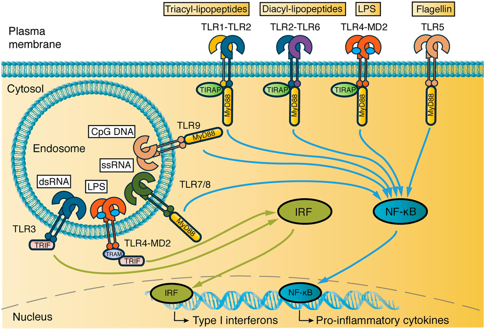
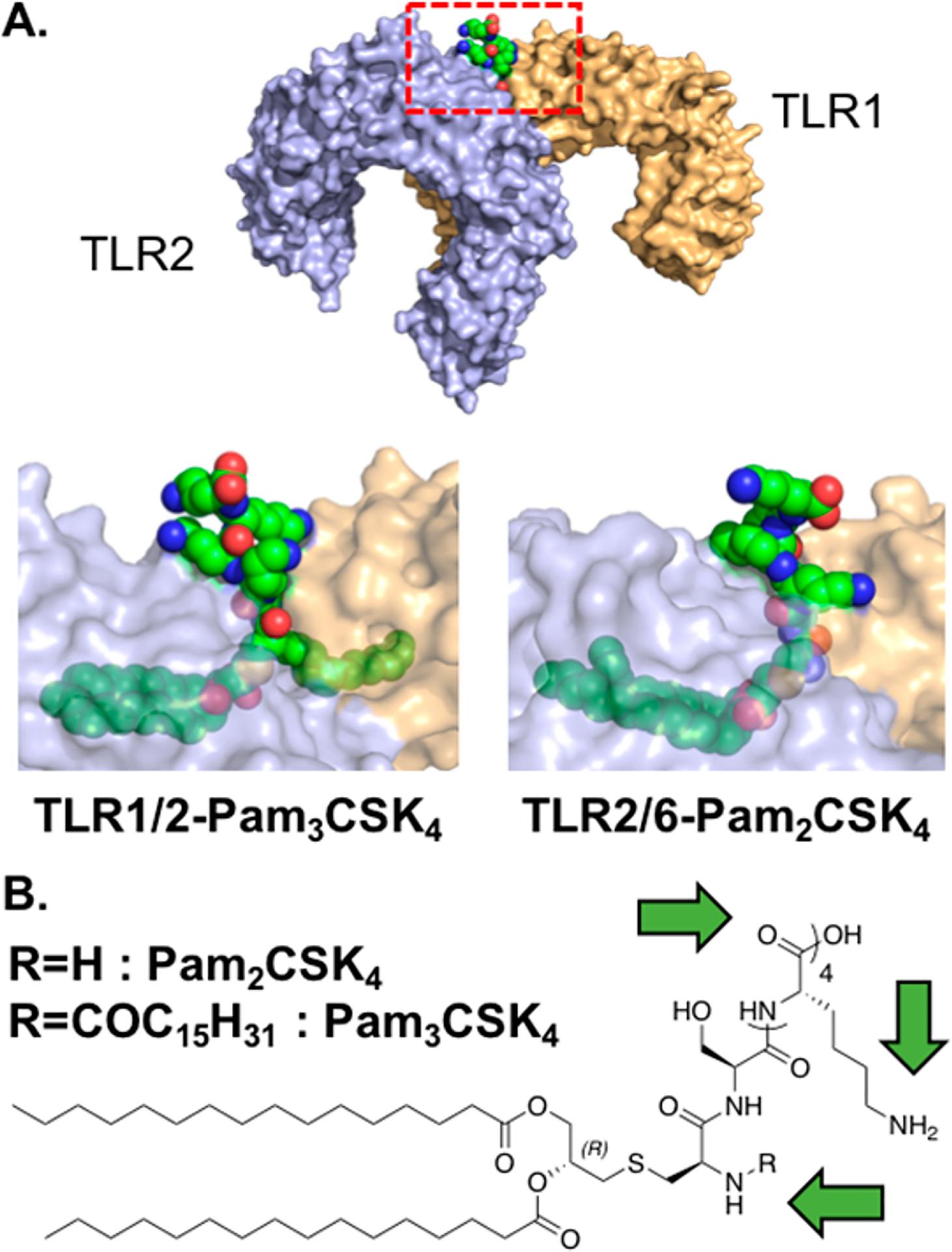
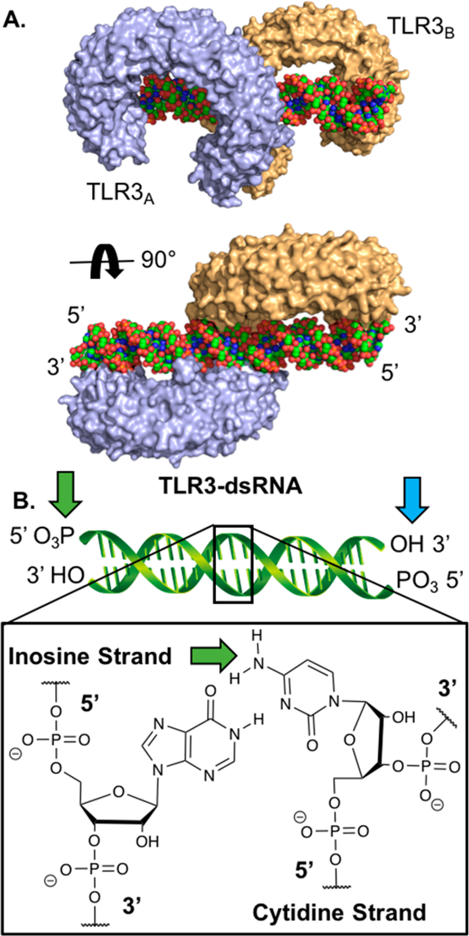
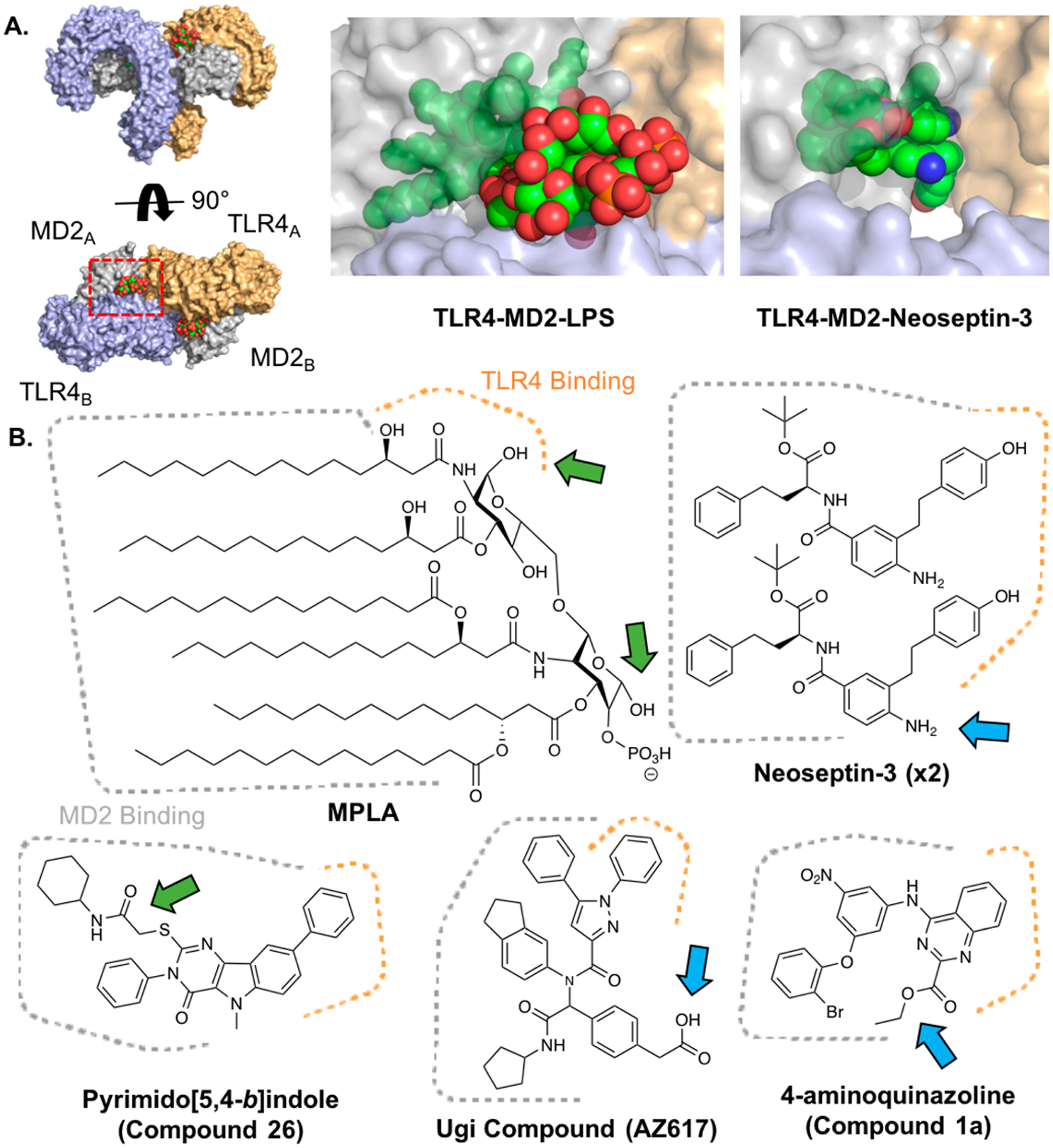
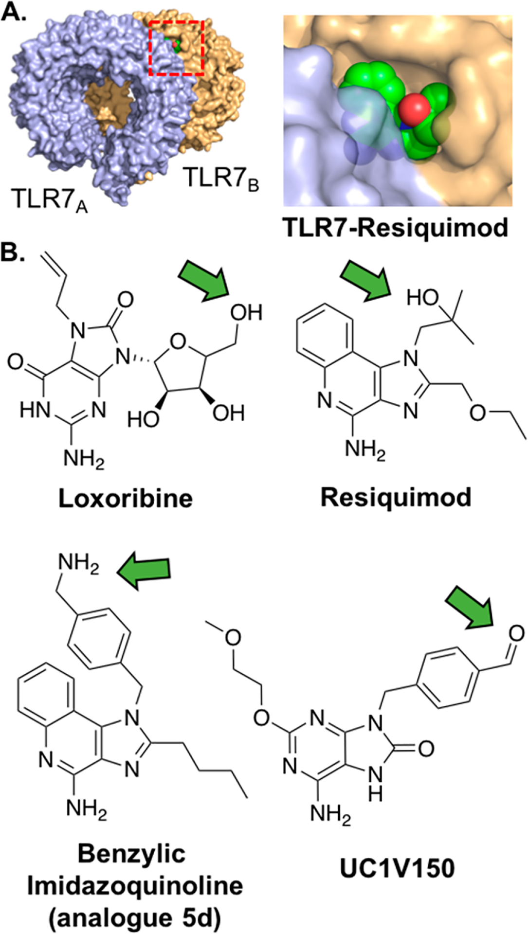
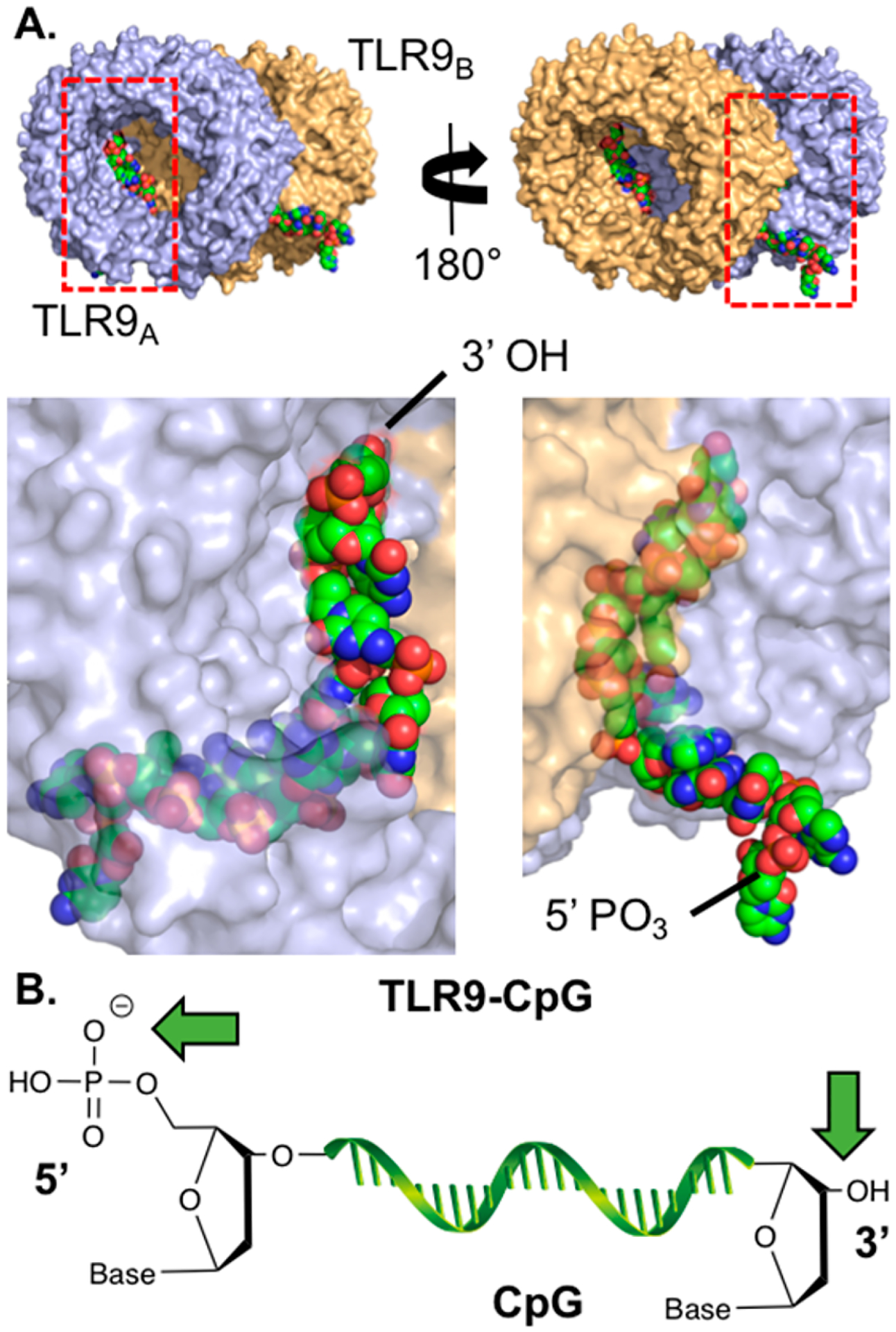
Toll-like receptors (TLRs) are vital elements of the mammalian immune system that function by recognizing pathogen-associated molecular patterns (PAMPs), bridging innate and adaptive immunity. They have become a prominent therapeutic target for the treatment of infectious diseases, cancer, and allergies, with many TLR agonists currently in clinical trials or approved as immunostimulants. Numerous studies have shown that conjugation of TLR agonists to other molecules can beneficially influence their potency, toxicity, pharmacokinetics, or function. The functional properties of TLR agonist conjugates, however, are highly dependent on the ligation strategy employed. Here, we review the chemical structural requirements for effective functional TLR agonist conjugation. In addition, we provide similar analysis for those that have yet to be conjugated. Moreover, we discuss applications of covalent TLR agonist conjugation and their implications for clinical use.


Conflict of interest statement

The authors declare no competing financial interest.

Figures

Figure 1.
Figure 1.
Cellular location of TLRs, their ligands, and signal transduction pathways. TLRs 1, 2, 6, 4, and 5 are located on the plasma membrane and signal, together with endosomal TLRs 7, 8, and 9, in a MyD88-dependent manner. The MyD88 pathway leads to NF-κB translocation to the nucleus and, ultimately, production of inflammatory cytokines. Activated TLR4 is endocytosed and, together with endosomal TLR3, signals through the IRF pathway, leading to Type I interferon production. MD2, TRAM, and TIRAP are adaptor proteins.
Figure 2.
Figure 2.
TLR2 agonists bind dimers of either TLR2-TLR1 or TLR2-TLR6. (A) Crystal structure of human TLR2-TLR1-Pam3CSK4 (PDB: 2Z7X). Red rectangle represents the area of the zoomed view of Pam3CSK4 binding to TLR2-TLR1. The analogous view from the TLR2-TLR6-Pam2CSK4 crystal structure is also shown (PDB: 5IJC). In both cases, the peptide C-terminus is solvent exposed. TLR2 is shown in blue, TLR1 and TLR6 are in orange, and lipopeptides are in green. TLRs in the zoomed view are shown at 40% transparency. (B) Chemical structure of Pam2CSK4 and Pam3CSK4. Green arrows highlight sites of conjugation.
Figure 3.
Figure 3.
TLR3 agonists are dsRNAs that bind to TLR3 dimers. (A) Crystal structure of mouse TLR3-dsRNA (PDB: 3CIY). The receptors bind to the negatively charged backbone of the dsRNA molecule. TLR3 is shown in blue and orange, and dsRNA is shown in green. (B) Carton structure of dsRNA with sites that have been conjugated highlighted by green arrows and suggested sites of conjugation by blue arrows.
Figure 4.
Figure 4.
TLR4 agonists bind MD2 and TLR4. (A) Crystal structure of human TLR4-MD2-LPS (PDB: 3FXI) from two viewpoints (left), red rectangle depicts zoom view of LPS binding (middle) in which proteins are shown with 40% transparency. The analogous view from the mouse TLR4-MD2-Neoseptin-3 crystal structure is also shown (PDB: 5IJC, right). TLR4 is shown in blue and orange, MD2 is shown in gray, and LPS and Neoseptin-3 are shown in green. (B) Chemical structures of various TLR4 agonists. Areas that bind MD2 (dotted gray) and TLR4 (dotted orange) shown. Green arrows highlight previous sites of conjugation; blue arrows highlight suggested sites of conjugation.
Figure 5.
Figure 5.
TLR7/8 agonists bind TLR7 and/or TLR8 homodimers. (A) Crystal structure of monkey TLR7-Resiquimod (PDB: 3FXI), red rectangle depicts view of Resiquimod binding (right) in which proteins are shown at 20% transparency. The molecule bridges the receptors at the interface. TLR7 is shown in blue and orange, and Resiquimod is shown in green. (B) Chemical structures of various TLR7/8 agonists. Green arrows highlight recommended sites of conjugation.
Figure 6.
Figure 6.
TLR9 agonists are ssDNAs that bind TLR9 dimers. (A) Two views of the horse TLR9-ssDNA (CpG 1668) crystal structure (PDB: 3WPC). Shown below each view is the zoomed region depicted by the red rectangle where TLR9 is shown at 40% transparency. The receptors bind to the CpG backbone and several nucleobases of the molecule. TLR9 is shown in blue and orange, and ssDNA is shown in green. (B) Carton structure of CpG. Green arrows highlight sites of conjugation.

References

    1. Kawai T, and Akira S (2010) The role of pattern-recognition receptors in innate immunity: update on Toll-like receptors. Nat. Immunol 11, 373–384. - PubMed
    1. Baldridge JR, McGowan P, Evans JT, Cluff C, Mossman S, Johnson D, and Persing D (2004) Taking a Toll on human disease: Toll-like receptor 4 agonists as vaccine adjuvants and monotherapeutic agents. Expert Opin. Biol. Ther 4, 1129–1138. - PubMed
    1. Schön MP, and Schön M (2008) TLR7 and TLR8 as targets in cancer therapy. Oncogene 27, 190–199. - PubMed
    1. Marshak-Rothstein A (2006) Toll-like receptors in systemic autoimmune disease. Nat. Rev. Immunol 6, 823. - PMC - PubMed
    1. Kanzler H, Barrat FJ, Hessel EM, and Coffman RL (2007) Therapeutic targeting of innate immunity with Toll-like receptor agonists and antagonists. Nat. Med 13, 552–559. - PubMed

Publication types

MeSH terms