Comparative Study
. 2018 Apr;188(4):904-915.
doi: 10.1016/j.ajpath.2017.12.019. Epub 2018 Jan 31.

Comparative Histopathologic Lesions of the Male Reproductive Tract during Acute Infection of Zika Virus in AG129 and Ifnar-/- Mice

Affiliations
Comparative Study

Comparative Histopathologic Lesions of the Male Reproductive Tract during Acute Infection of Zika Virus in AG129 and Ifnar-/- Mice

Chad S Clancy et al. Am J Pathol. 2018 Apr.

Abstract

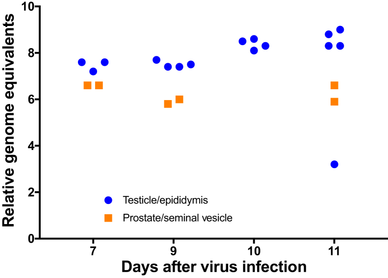
An understanding of the pathogenesis of infection with the Zika virus in the male reproductive tract is vital for the development of vaccines and antivirals that will limit or prevent sexual transmission. Two common immunocompromised mouse strains used in transmission studies-male with genes encoding interferon types I and II receptor gene knockout (IFNAR/IFNGR; AG129) and with interferon type 1 receptor knockout (Ifnar-/-) were infected with a Puerto Rican Zika virus isolate (PRVABC59), and pathology was assessed 5 to 11 days after infection. Virus was detected by immunohistochemistry and quantitative RT-PCR in the testicle and epididymis of AG129 and Ifnar-/- mice, and by immunohistochemistry in the prostate and seminal vesicle of infected AG129 mice. Severe disease manifestations initiating as epididymitis and progressing to orchitis were observed in both models, with more severe inflammation noted in the AG129 mouse strain. Significant inflammation was not observed in any evaluated accessory sex gland at any point during infection. Time-course analysis of infection revealed an increase in the severity of disease within the epididymis of both strains, indicating a potential route of sexual transmission. Male mice with Ifnar-/- may better recapitulate Zika virus in humans and provide insight into the mechanism of sexual transmission, due to milder histopathologic lesions, the presence of histologically normal sperm in epididymal tubules, and an ability to survive the acute phase of disease.


Figures

Figure 1
Figure 1
A: Interferon type I and II receptor gene knockout (AG129) control testicle. Seminiferous tubules are supported by a distinct basement membrane (black arrow) and lined by Sertoli cells and spermatogonia. Within the lumen of each tubule are moderate numbers to numerous maturing spermatozoa (arrowhead). The interstitium contains small blood vessels, which are abutted by Leydig (interstitial) cells (white arrow). Hematoxylin and eosin (H&E) stain. B: AG129 testicle, 9 days after infection (DPI). The interstitium is infiltrated by neutrophils (arrow). There is loss of orderly maturation and necrosis of spermatids, and necrosis of tubular cells (arrowhead). H&E stain. C: Interferon type I receptor gene knockout (Ifnar−/−) testicle, 8 DPI. The interstitium is multifocally infiltrated and expanded by low numbers of neutrophils (arrow). Seminiferous tubules contain abundant maturing spermatids and spermatozoa. H&E stain. D: AG129 control epididymis. Tubules are lined by a single layer of tall, columnar epithelial cells with stereocilia, and tubular lumens contain numerous maturing spermatids (arrow). The interstitial spaces are populated by low numbers of fibroblasts and rare lymphocytes within a fine fibrovascular stroma. H&E stain. E: AG129 epididymis, 7 DPI. Variable numbers of tubular epithelial cells are undergoing necrosis (arrow). Diffusely, tubules contain necrotic epithelial cells admixed with maturing spermatozoa (arrowhead). H&E stain. F:Ifnar−/− epididymis, 8 DPI. Low numbers of tubular epithelial cells undergo single-cell necrosis (arrow) and slough into the tubular lumen. Tubules contain abundant maturing spermatozoa. The interstitium is multifocally infiltrated by low to moderate numbers of inflammatory cells, predominantly lymphocytes and fewer neutrophils (arrowhead). H&E stain. Original magnification: ×20 (A, D); ×40 (B, C, E, F). Scale bars: 100 μm (A, D); 50 μm (B, C, E, F).
Figure 2
Figure 2
A and B: Histopathologic lesion severity in the reproductive tract of interferon type I and II receptor gene knockout (AG129) and interferon type I receptor gene knockout (Ifnar−/−) mice. AG129 mice exhibit increasing severity of orchitis (A) and epididymitis (B), with 100% exhibiting severe inflammation by 11 days after infection (DPI). C and D:Ifnar−/− mice exhibit an age-dependent disease phenotype, with 100% mortality in 3-week-old mice by 6 and 7 DPI, with 17% (n = 1) exhibiting moderate orchitis and severe epididymitis in 33% (n = 2) at the terminal phase of disease. C: In contrast, 6- and 16-week-old males exhibit no to mild orchitis at 5, 8, and 11 DPI. D: Epididymitis progresses from absent to mild at 5 DPI to minimal to moderate at 11 DPI in both 6- and 16-week-old Ifnar−/− mice. n = 5 (A and B); n = 6 (C and D).
Figure 3
Figure 3
A: Interferon type I receptor gene knockout (Ifnar−/−) preputial gland, 8 days after infection (DPI). The periglandular adipose and interstitial tissue is infiltrated by low to moderate numbers of neutrophils and lymphocytes (arrow). Preputial glands are composed of epithelial cells, which undergo holocrine secretion (arrowhead). Hematoxylin and eosin (H&E) stain. B:Ifnar−/− ventral prostate, 8 DPI. The lamina propria underlying the prostatic epithelium is infiltrated by low to moderate numbers of neutrophils (arrow). Low numbers of prostatic epithelial cells are necrotic and slough into the prostatic lumen (arrowhead). The lumen contains moderate numbers of sperm (retrograde movement, handling artefact). H&E stain. C:Ifnar−/− vas deferens, 8 DPI. The entire vas deferens is lined by pseudociliated epithelial cells that show immunoreactivity for Zika virus antigen. Polyclonal anti-Zika antibody; counterstain, hematoxylin. D:Ifnar−/− preputial gland, 8 DPI. Preputial gland epithelial cells show variable immunoreactivity to Zika virus antigen. Polyclonal anti-Zika antibody; counterstain hematoxylin. E:Ifnar−/− ventral prostate, 8 DPI. Moderate numbers of prostatic epithelial cells and necrotic epithelial cells are immunoreactive to Zika virus antigen. Polyclonal anti-Zika antibody; counterstain, hematoxylin. F:Ifnar−/− ampullary gland, 8 DPI. The ampullary gland (right) is an outpouching of the vas deferens (left) and is multifocally lined by epithelial cells that show immunoreactivity to Zika virus antibody. Polyclonal anti-Zika antibody, counterstain, hematoxylin. All IHC images were pseudocolored utilizing Photoshop (Adobe Systems, San Jose, CA) to convert the diaminobenzidine peroxidase brown chromogen to red to highlight the immunoreactivity to the hematoxylin counterstain. Original magnification: ×40 (A and B); ×20 (C–F). Scale bars: 50 μm (A and B); 100 μm (C–F).
Figure 4
Figure 4
A: Interferon type I receptor gene knockout (Ifnar)−/− control testicle. Polyclonal anti-Zika antibody, counterstain, hematoxylin. B: Interferon type I and II receptor gene knockout (AG129) testicle, 10 days after infection (DPI). Seminiferous tubules are lined by variable numbers of cells that exhibit immunoreactivity to Zika virus. Polyclonal anti-Zika antibody, counterstain, hematoxylin. C:Ifnar−/− testicle, 11 DPI. Low numbers of seminiferous tubules are partially lined by cells that exhibit immunoreactivity to Zika virus antibody. Polyclonal anti-Zika antibody; counterstain, hematoxylin. D:Ifnar−/− control epididymis. Polyclonal anti-Zika antibody, counterstain, hematoxylin. E: AG129 epididymis, 5 DPI. Within the head of the epididymis, most tubules are lined by moderate numbers of epithelial cells that show immunoreactivity to Zika virus antibody. Polyclonal anti-Zika antibody, counterstain, hematoxylin. F:Ifnar−/− epididymis, 11 DPI. Within the head of the epididymis, most tubules are lined by low to moderate numbers of epithelial cells that show immunoreactivity to Zika virus antibody. Polyclonal anti-Zika antibody; counterstain, hematoxylin. All IHC images were pseudocolored utilizing Photoshop (Adobe Systems, San Jose, CA) to convert the diaminobenzidine peroxidase brown chromogen to red to highlight the immunoreactivity to the hematoxylin counterstain. Original magnification, ×20. Scale bars = 100 μm.
Figure 5
Figure 5
ZIKV RNA in reproductive tissue in interferon type I and II receptor gene knockout (AG129) males. ZIKV RNA remains at relatively constant levels in both the epididymis and testicle of 6-week-old AG129 males from 7 to 11 days after infection (DPI). ZIKV is detectable by quantitative RT-PCR in pooled homogenates of the prostate and seminal vesicle from 7 to 11 DPI.
Figure 6
Figure 6
A and B: ZIKV RNA in the reproductive tissue of interferon type I receptor gene knockout mice. ZIKV is detectable in both the testicle (A) and epididymis (B) of infected males at 5 to 11 days after infection. GE, genome equivalents.

References

    1. Dick G.W. Zika virus (I). Isolations and serological specificity. Trans R Soc Trop Med Hyg. 1952;46:509–520. - PubMed
    1. Roth A., Mercier A., Lepers C., Hoy D., Duituturaga S., Benyon E., Guillaumot L., Souarès Y. Concurrent outbreaks of dengue, chikungunya and Zika virus infections—an unprecedented epidemic wave of mosquito-borne viruses in the Pacific 2012-2014. Euro Surveill. 2014;19 pii:20929. - PubMed
    1. Heang V., Yasuda C.Y., Sovann L., Haddow A.D., da Rosa A.P.T., Tesh R.B., Kasper M.R. Zika virus infection, Cambodia, 2010. Emerg Infect Dis. 2012;18:349–351. - PMC - PubMed
    1. Duffy M.R., Chen T.H., Hancock W.T., Powers A.M., Kool J.L., Lanciotti R.S., Pretrick M., Marfel M., Holzbauer S., Dubray C., Guillaumot L., Griggs A., Bel M., Lambert A.J., Laven J., Kosoy O., Panella A., Biggerstaff B.J., Fischer M., Hayes E.B. Zika virus outbreak on Yap Island, Federated States of Micronesia. N Engl J Med. 2009;360:2536–2543. - PubMed
    1. Yockey L.J., Varela L., Rakib T., Khoury-Hanold W., Fink S.L., Stutz B., Szigeti-Buck K., Van den Pol A., Lindenbach B.D., Horvath T.L., Iwasaki A. Vaginal exposure to Zika virus during pregnancy leads to fetal brain infection. Cell. 2016;166:1247–1256.e4. - PMC - PubMed

Publication types