. 2018 May 1:287:31-41.
doi: 10.1016/j.toxlet.2018.01.017. Epub 2018 Jan 31.

Alterations in the nigrostriatal dopamine system after acute systemic PhIP exposure

Affiliations

Alterations in the nigrostriatal dopamine system after acute systemic PhIP exposure

Zeynep Sena Agim et al. Toxicol Lett. .

Abstract

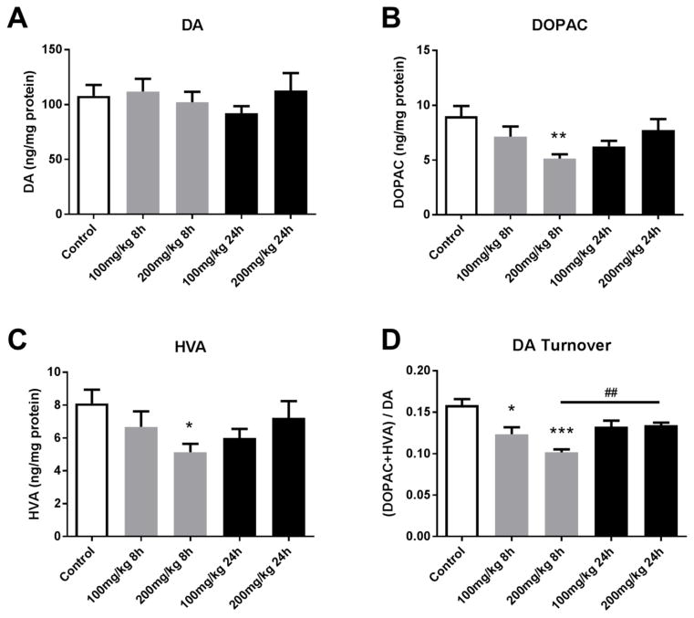
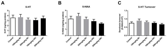
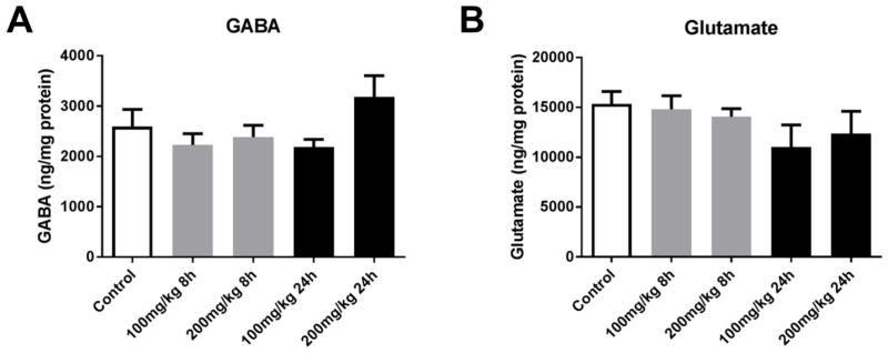
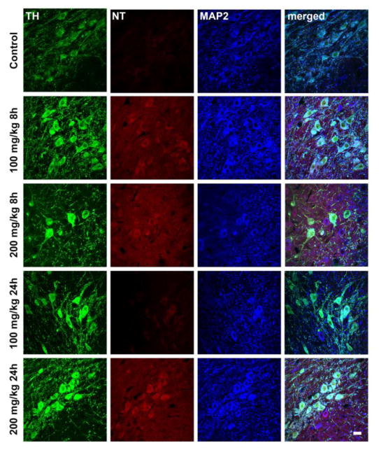
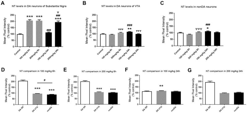
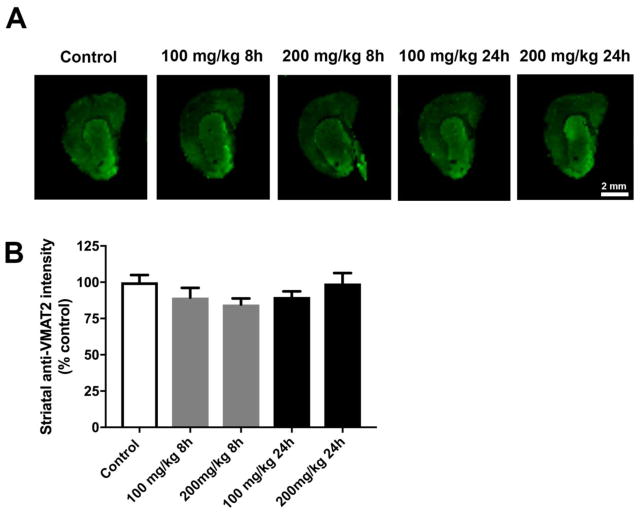
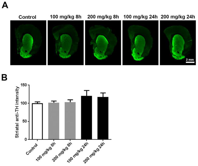
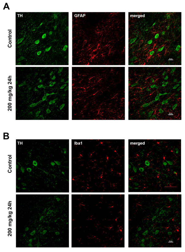
Heterocyclic amines (HCAs) are primarily formed during cooking of meat at high temperature. HCAs have been extensively studied as mutagens and possible carcinogens. Emerging data suggest that HCAs are neurotoxic and may be relevant to Parkinson's disease (PD) etiology. However, the majority of HCAs have not been evaluated for in vivo neurotoxicity. Here, we investigated acute in vivo neurotoxicity of 2-amino-1-methyl-6-phenylimidazo[4,5-b]pyridine (PhIP). PhIP is the most prevalent genotoxin in many types of meats. Adult, male Sprague-Dawley rats were subjected to acute, systemic PhIP at doses and time-points that have been extensively utilized in cancer studies (100 and 200 mg/kg for 8, 24 h) and evaluated for changes in dopaminergic, serotoninergic, GABAergic, and glutamatergic neurotransmission. PhIP exposure resulted in decreased striatal dopamine metabolite levels and dopamine turnover in the absence of changes to vesicular monoamine transporter 2 levels; other neurotransmitter systems were unaffected. Quantification of intracellular nitrotyrosine revealed higher levels of oxidative damage in dopaminergic neurons in the substantia nigra after PhIP exposure, while other neuronal populations were less sensitive. These changes occurred in the absence of an overt lesion to the nigrostriatal dopamine system. Collectively, our study suggests that acute PhIP treatment in vivo targets the nigrostriatal dopaminergic system and that PhIP should be further examined in chronic, low-dose studies for PD relevance.

Keywords: Dopaminergic toxicity; Heterocyclic amines; Oxidative stress; Parkinson’s disease; PhIP.


Figures

Figure 1
Figure 1. Acute PhIP treatment decreases striatal dopamine metabolite levels and dopamine turnover
Animals received a single oral dose of PhIP (100 or 200 mg/kg) and samples were obtained 8 or 24 h later. Striatal dopamine (DA) (A), 3,4-dihydroxyphenylacetic acid (DOPAC) (B) and homovanillic acid (HVA) (C) levels were measured using HPLC with electrochemical detection. Neurotransmitter levels were normalized to total tissue protein and expressed as ng neurotransmitter/mg protein. Dopamine turnover (D) was determined as (DOPAC + HVA)/DA. *p< 0.05, **p< 0.01, ***p<0.001 vs. control; ##p<0.01 8 h vs. 24 h (200 mg/kg); Kruskal-Wallis test, followed by Dunn’s multiple comparisons post hoc test. The data are presented as the mean ± SEM (n = 8–10/treatment).
Figure 2
Figure 2. Acute PhIP treatment does not alter striatal serotonin or metabolite levels
Animals received a single oral dose of PhIP (100 or 200 mg/kg) and samples were obtained 8 or 24 h later. Striatal serotonin (5-HT) (A) and 5-hydroxyindoleacetic acid (5-HIAA) (B) levels were measured using HPLC with electrochemical detection. Neurotransmitter levels were normalized to total tissue protein and expressed as ng neurotransmitter/mg protein. 5-HT turnover (C) was determined as 5-HIAA/5-HT. The data are presented as the mean ± SEM (n = 8–10/treatment).
Figure 3
Figure 3. Acute PhIP treatment does not alter amino acid neurotransmitter levels
Animals received a single oral dose of PhIP (100 or 200 mg/kg) and samples were obtained 8 or 24 h later. Striatal gamma-aminobutyric acid (GABA) (A) and glutamate (B) levels were measured using HPLC with electrochemical detection. Neurotransmitter levels were normalized to total tissue protein and expressed as ng neurotransmitter/mg protein. The data are presented as the mean ± SEM (n = 8–10/treatment).
Figure 4
Figure 4. Acute PhIP treatment increases histological evidence of oxidative damage in dopaminergic neurons of substantia nigra
Animals received a single oral dose of PhIP (100 or 200 mg/kg) and samples were obtained 8 or 24 h later. Coronal midbrain sections from control and treatment groups were stained for tyrosine hydroxylase (TH) (in green), nitrotyrosine (NT) (in red) and microtubule-associated protein 2 (MAP2) (in blue). Scale bars = 20 μm.
Figure 5
Figure 5. Quantitative analysis of cellular nitrotyrosine levels shows that PhIP treatment produces oxidative damage in dopaminergic neurons of substantia nigra relative to other cell populations
To quantify oxidative damage, ROIs surrounding cell bodies of dopaminergic (DA) neurons (TH+) in substantia nigra (SN) were drawn and NT intensity in each ROI was quantified, and normalized to the mean of the control (A). The data are presented as the mean ± SEM (n = 390–671 cell bodies from 5 animals/treatment). NT levels were also quantified in DA neurons from the ventral tegmental area (VTA) (B) and non-dopaminergic (nonDA) neurons localized dorsally to the SN (C) (n = 162–287 bodies from 3 animals/treatment and n = 129–325 cell bodies from 3 animals/treatment, respectively). *p<0.05, **p<0.01, ***p<0.001 compared to control; ###p<0.001 compared to same-dose group; Ψp<0.05, ΨΨΨp<0.001 compared to same-time group; Kruskal-Wallis test followed by Dunn’s multiple comparisons post hoc test. To directly compare the magnitude of NT level changes for each of the three cell populations, normalized values were analyzed separately for each dose and time-point (D–G). **p<0.01, ***p<0.001 compared to DA SN; #p<0.05 compared to indicated group; Kruskal-Wallis test followed by Dunn’s multiple comparisons post hoc test.
Figure 6
Figure 6. Acute PhIP treatment does alter striatal VMAT2 levels
Coronal sections from the striatum from control and PhIP-treated animals were immunofluorescently stained for VMAT2 (A). Immunofluorescent intensity of ROIs surrounding the striatum was quantified using Image Studio version 3.1. All quantifications were normalized to average of control group (B). Data are presented as mean ± SEM (n = 4 or 5 sections/animal from 5 animals per treatment).
Figure 7
Figure 7. Acute PhIP treatment does not cause striatal terminal loss
Coronal sections from the striatum from control and PhIP-treated animals were immunofluorescently stained for tyrosine hydroxylase (TH) (A). Immunofluorescent intensity of ROIs surrounding the striatum was quantified using Image Studio version 3.1. All quantifications were normalized to average of control group (B). Data are presented as mean ± SEM (n = 4 or 5 sections/animal from 10 animals per treatment).
Figure 8
Figure 8. Acute PhIP exposure does not induce overt neuroinflammation in the substantia nigra
Sections from the substantia nigra from control and 200 mg/kg for 24h groups were stained for TH (in green) and GFAP (in red) (A). The nigral sections from control and 200 mg/kg for 24h group were stained for TH (in green) and Iba1 (in red) (B). Scale bars represent 20 μm.

References

    1. Augustsson K, Skog K, Jagerstad M, Steineck G. Assessment of the human exposure to heterocyclic amines. Carcinogenesis. 1997;18(10):1931–5. - PubMed
    1. Burke WJ, et al. Aggregation of alpha-synuclein by DOPAL, the monoamine oxidase metabolite of dopamine. Acta Neuropathol. 2008;115(2):193–203. doi: 10.1007/s00401-007-0303-9. - DOI - PubMed
    1. Byrne C, Sinha R, Platz EA, Giovannucci E, Colditz GA, Hunter DJ, Speizer FE, Willett WC. Predictors of dietary heterocyclic amine intake in three prospective cohorts. Cancer epidemiology, biomarkers & prevention: a publication of the American Association for Cancer Research, cosponsored by the American Society of Preventive Oncology. 1998;7(6):523–9. - PubMed
    1. Cai Z. Monoamine oxidase inhibitors: promising therapeutic agents for Alzheimer’s disease (Review) Mol Med Rep. 2014;9(5):1533–41. doi: 10.3892/mmr.2014.2040. - DOI - PubMed
    1. Cannon JR, Greenamyre JT. Neurotoxic in vivo models of Parkinson’s disease recent advances. Prog Brain Res. 2010;184:17–33. doi: 10.1016/S0079-6123(10)84002-6. - DOI - PubMed

MeSH terms

LinkOut - more resources