. 2018 Nov;25(1):388-397.
doi: 10.1080/10717544.2018.1431979.

Development a hyaluronic acid ion-pairing liposomal nanoparticle for enhancing anti-glioma efficacy by modulating glioma microenvironment

Affiliations

Development a hyaluronic acid ion-pairing liposomal nanoparticle for enhancing anti-glioma efficacy by modulating glioma microenvironment

Liuqing Yang et al. Drug Deliv. 2018 Nov.

Abstract

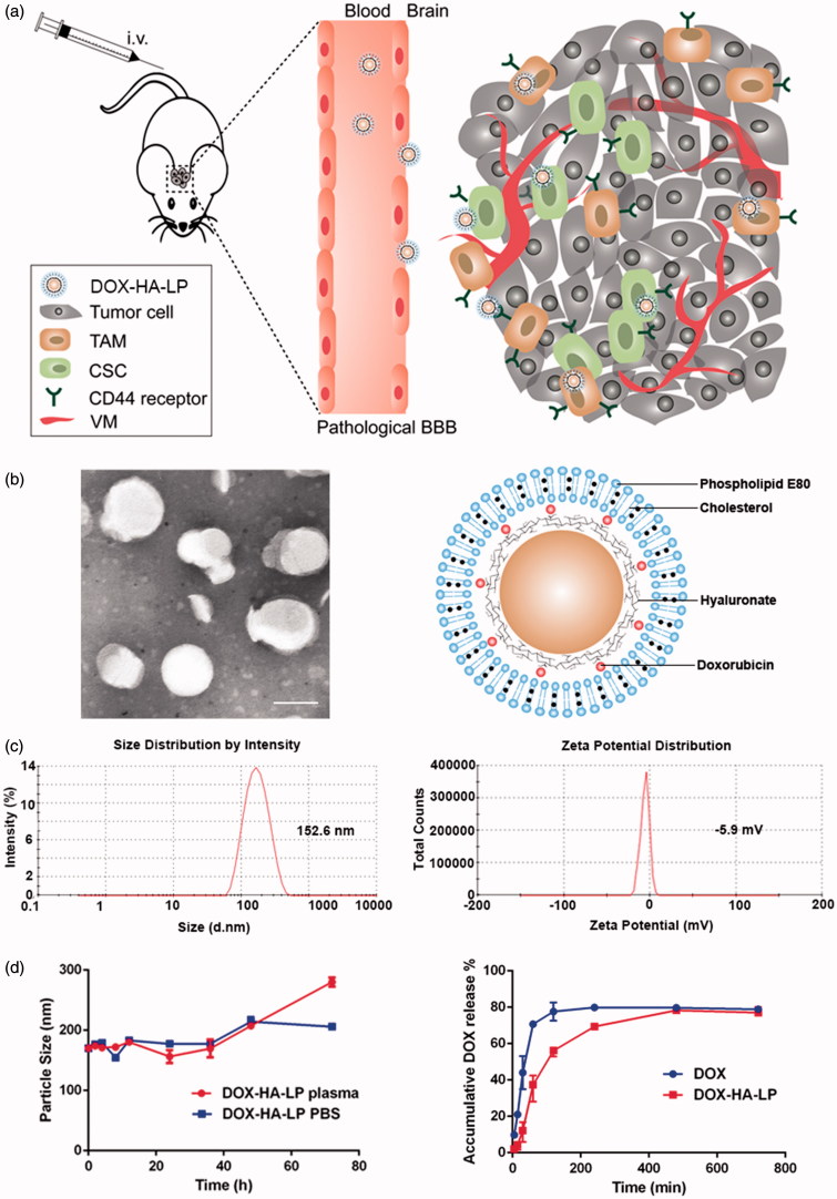
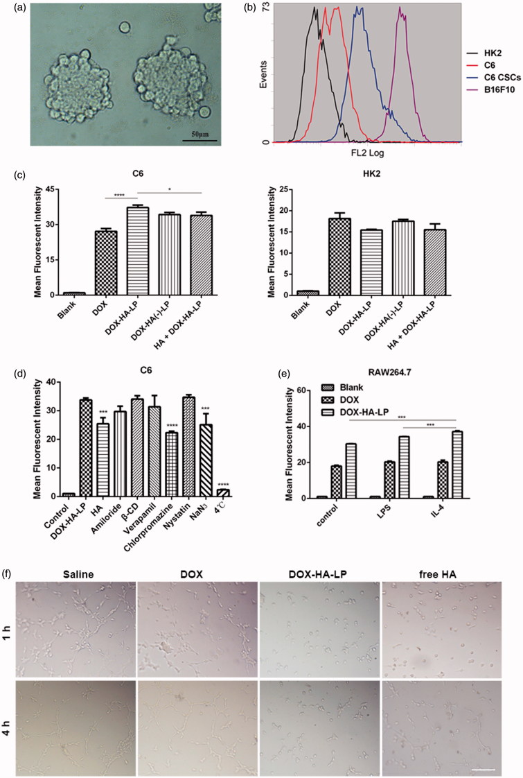
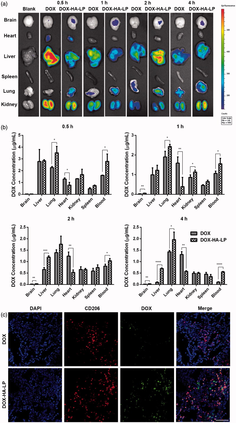
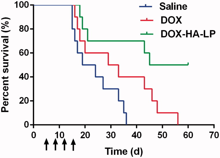
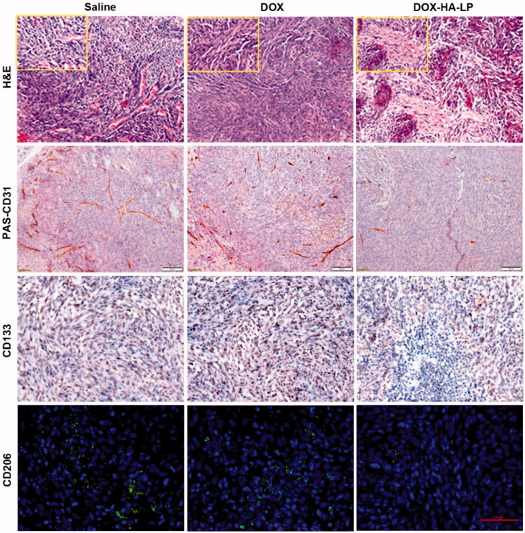
Glioma, one of the most common brain tumors, remains a challenge worldwide. Due to the specific biological barriers such as blood-brain barrier (BBB), cancer stem cells (CSCs), tumor associated macrophages (TAMs), and vasculogenic mimicry channels (VMs), a novel versatile targeting delivery for anti-glioma is in urgent need. Here, we designed a hyaluronic acid (HA) ion-pairing nanoparticle. Then, these nanoparticles were encapsulated in liposomes, termed as DOX-HA-LPs, which showed near-spherical morphology with an average size of 155.8 nm and uniform distribution (PDI = 0.155). HA was proven to specifically bind to CD44 receptor, which is over-expressed on the surface of tumor cells, other associated cells (such as CSCs and TAMs) and VMs. We systematically investigated anti-glioma efficacy and mechanisms in vivo and in vitro. The strong anti-glioma efficacy could attribute to the accumulation in glioma site and the regulation of tumor microenvironment with depletion of TAMs, inhibition of VMs, and elimination of CSCs.

Keywords: CD44; CSCs; Hyaluronic acid; TAMs; glioma.


Figures

Figure 1.
Figure 1.
Characterization of DOX-HA-LPs. (a) Schematic illustration of DOX-HA-LPs, which is transported into glioma site after tail vein injection, targeting TAMs and CSCs mediated by CD44, resulting in depletion of TAMs, inhibition of VMs and elimination of CSCs; (b) TEM image and schematic diagrams of DOX-HA-LPs. Scale bar, 100 nm; (c) DLS size and zeta-potential measurement of DOX-HA-LPs; (d) the stability of DOX formulation in plasma and PBS for 72 h; DOX release profiles of DOX formulations at pH 7.4 (n = 3, mean ± SD).
Figure 2.
Figure 2.
The effect of different formulations on cells. (a) Microscopic image of C6 CSCs spheres. Scale bar, 50 μm; (b) expression of CD44 at C6, C6 CSCs, B16F10 (as a positive control cell line) and HK2 (as a negative control cell line) cells; (c) cellular uptake of DOX formulations after incubation with HK2, C6 cells for 4 h. HA + DOX-HA: cells were incubated with HA solution (10 mg/mL, 100 kDa) in advance. Data represent mean ± SD (n = 3). *p < .05, **p < .01, ****p < .0001; (d) endocytosis inhibition assay on C6 cells. Data represent mean ± SD (n = 3). ***p < .001. ****p < .0001; (e) DOX-HA-LPs selectively targeted IL-4 treated RAW 264.7. Data represent mean ± SD (n = 3). ***p < .001; (f) the destruction of VMs channels after treatment with different DOX formulations. Scale bar, 100 μm.
Figure 3.
Figure 3.
DOX-HA-LPs exhibit a different circulating behavior and a glioma-bearing brains concentrated capability. (a) Ex vivo images showed the drug biodistribution of DOX formulations in C6 glioma-bearing mice at 0.5, 1, 2, and 4 h after intravenous administration; (b) the mean concentrations of DOX in different tissues of C6 glioma-bearing mice after intravenous administration of DOX formulations at 0.5, 1, 2, and 4 h. Data represent mean ± SD (n = 5); (c) in vivo fluorescent microscopy images of brain sections from C6 glioma-bearing mice at 1 h post-injection of DOX and DOX-HA-LPs. TAMs were identified by CD206 marker and nuclei stained with DAPI. Scale bar, 50 μm.
Figure 4.
Figure 4.
The survival curves of C6 glioma-bearing mice treated with DOX formulations (2 mg/kg DOX) at day 5, 8, 11, and 14 after inoculation. Data represent mean ± SD (n = 10).
Figure 5.
Figure 5.
H&E staining of brain glioma site at 24 h after antitumor dosage administration of saline, free DOX and DOX-HA-LPs. Scale bar, 50 μm and 20 μm (small boxes). CD34-PAS dual staining, CD133 staining, CD206 staining on the brain glioma site. Scale bar, 50 μm.

References

    1. Allen TM. (1994). The use of glycolipids and hydrophilic polymers in avoiding rapid uptake of liposomes by the mononuclear phagocyte system. Adv Drug Deliv Rev 13:285–309.
    1. Cohen ZR, Ramishetti S, Peshes-Yaloz N, et al. (2015). Localized RNAi therapeutics of chemoresistant grade iv glioma using hyaluronan-grafted lipid-based nanoparticles. ACS Nano 9:1581–91. - PubMed
    1. Hao NB, Lu MH, Fan YH, et al. (2012). Macrophages in tumor microenvironments and the progression of tumors. Clin Dev Immunol 2012:948098. - PMC - PubMed
    1. Hayward SL, Wilson CL, Kidambi S. (2016). Hyaluronic acid-conjugated liposome nanoparticles for targeted delivery to CD44 overexpressing glioblastoma cells. Oncotarget 7:34158–71. - PMC - PubMed
    1. Hu CMJ, Zhang L, Aryal S, et al. (2011). Erythrocyte membrane-camouflaged polymeric nanoparticles as a biomimetic delivery platform. Proc Natl Acad Sci USA 108:10980–5. - PMC - PubMed