. 2018 Jan 29;7(3):e007197.
doi: 10.1161/JAHA.117.007197.

Neuronal PirB Upregulated in Cerebral Ischemia Acts as an Attractive Theranostic Target for Ischemic Stroke

Affiliations

Neuronal PirB Upregulated in Cerebral Ischemia Acts as an Attractive Theranostic Target for Ischemic Stroke

Jie Wang et al. J Am Heart Assoc. .

Abstract

Background: Ischemic stroke is a complex disease with multiple etiologies and clinical manifestations. Paired immunoglobulin-like receptor B (PirB), which is originally thought to function exclusively in the immune system, is now also known to be expressed by neurons. A growing number of studies indicate that PirB can inhibit neurite outgrowth and restrict neuronal plasticity. The aim of the study is to investigate whether PirB can be an attractive theranostic target for ischemic stroke.

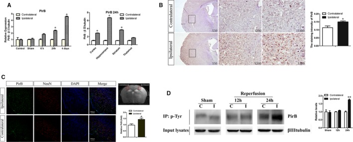
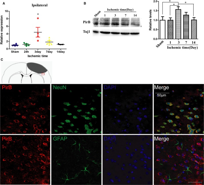
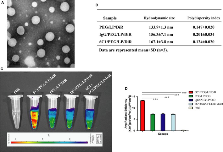
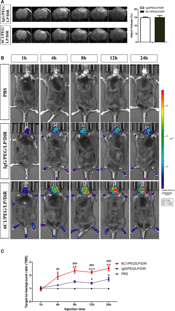
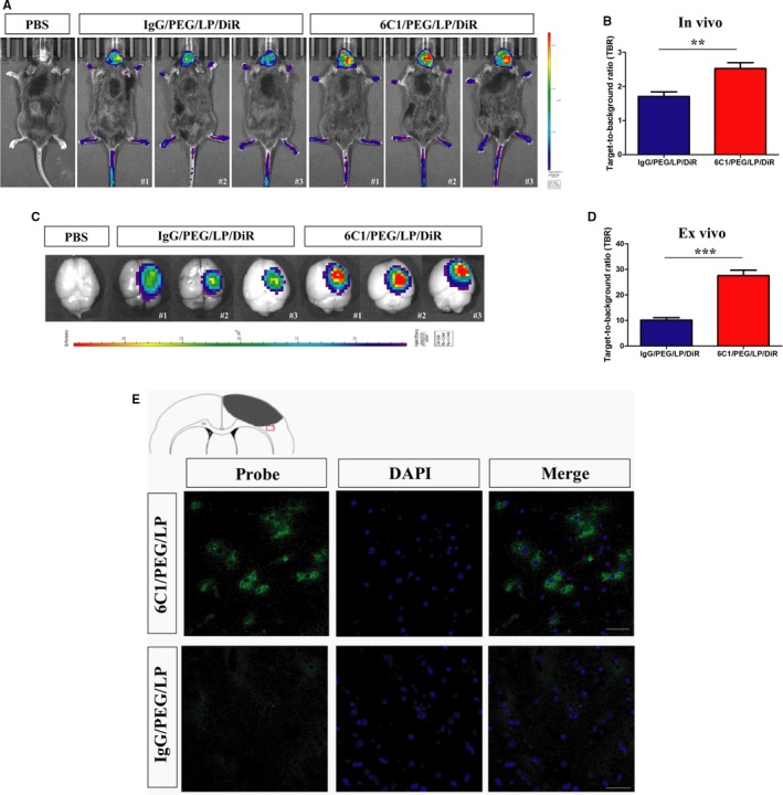
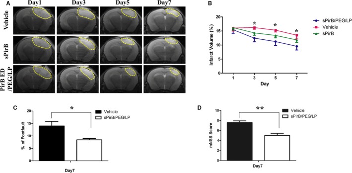
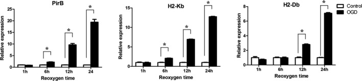
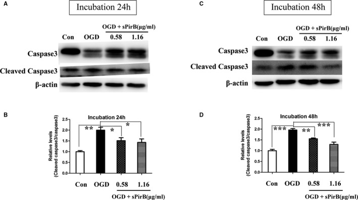
Methods and results: First, we investigated the spatial-temporal expression of PirB in multiple ischemic stroke models, including transient middle cerebral artery occlusion, photothrombotic cerebral cortex ischemia, and the neuronal oxygen glucose deprivation model. Then, anti-PirB immunoliposome nanoprobe was developed by thin-film hydration method and investigated its specific targeting in vitro and in vivo. Finally, soluble PirB ectodomain (sPirB) protein delivered by polyethylene glycol-modified nanoliposome was used as a therapeutic reagent for ischemic stroke by blocking PirB binding to its endogenous ligands. These results showed that PirB was significantly upregulated after cerebral ischemic injury in ischemic stroke models. Anti-PirB immunoliposome nanoprobe was successfully developed and specifically bound to PirB in vitro. There was accumulation of anti-PirB immunoliposome nanoprobe in the ischemic hemisphere in vivo. Soluble PirB ectodomains remarkably improved ischemic stroke model recovery by liposomal delivery system.

Conclusions: These data indicated that PirB was a significant element in the pathological process of cerebral ischemia. Therefore, PirB may act as a novel theranostic target for ischemic stroke.

Keywords: PirB; imaging; ischemic stroke; liposome; therapy.


Figures

Figure 1
Figure 1
A through D, Enhanced PirB expression and PirB signaling after transient middle cerebral artery occlusion (MCAO). A, Real‐time quantitative PCR analysis revealed highly increased PirB mRNA in the ischemic hemisphere compared with the undamaged contralateral side, sham‐operated hemisphere, and control group before MCAO both at 24 hours and at 4 days post‐MCAO (n=5). B, Representative images of immunohistochemical staining showed that PirB protein levels were markedly increased in the damaged hemisphere 24 hours post‐MCAO, compared with the undamaged contralateral side. Staining intensity of molecule was quantified by the simple PCI image analysis software (n=4). C, Majority of PirB immunostaining (green) colocalized with neuronal marker NeuN (red) in the cortical penumbra 24 hours after ischemia. Rectangle in T2‐weighted image indicates cortical area used for cell staining. Quantification of PirB staining in tMCAO brain slices was by ImageJ software (NIH, Bethesda, MD). Scale bar=100 μm (n=4). D, Protein extracts from hemispheres of post‐MCAO brains were subjected to immunoprecipitation for phosphotyrosine to assess levels of tyrosine phosphorylation of PirB. Expression of βIIItubulin was detected in input lysates. Phosphorylation of PirB was quantified as the ratio of band density to that of βIIItubulin. C indicates contralateral to ischemia; I, ipsilateral to ischemia; Sham, sham‐operated mice (n=4). Data are the mean±SEM. Statistical significance is indicated by: *P<0.05; **P<0.01. NeuN indicates neuronal nuclei; PirB, paired immunoglobulin‐like receptor B.
Figure 2
Figure 2
A through C, Expression and cellular location of PirB after cerebral cortex ischemia. Expression of PirB mRNA (A) and protein level (B) in the cortex with ischemia at different time points were detected by real‐time quantitative PCR and western blot analysis, respectively (n=4). Data are the mean±SEM. Statistical significance is indicated by *P<0.05, the expression level of ischemic cortex at different time points post‐injury compared with sham‐operated cortex. C, Representative images of PirB (red) and NeuN (green), or PirB (red) and GFAP (astrocytes, green) double immunofluorescene staining in the ischemic border zone of brain sections are presented. Scale bar=50 μm. GFAP indicates glial fibrillary acidic protein; NeuN, neuronal nuclei; PirB, paired immunoglobulin‐like receptor B; Tuj1, neuron‐specific Class III β‐tubulin.
Figure 3
Figure 3
A, Transmission electron microscopy image of 6C1/PEG/LP/DiR. B, Hydrodynamic size and polydispersity index of PEG/LP/DiR, IgG/PEG/LP/DiR, and 6C1/PEG/LP/DiR were determined by dynamic light scattering. C, Representative near‐infrared fluorescence (NIRF) images of spleen cells in different groups showed a remarkable fluorescence signal in the 6C1/PEG/LP/DiR group, which indicated a higher cellular binding efficacy of 6C1/PEG/LP/DiR compared with the control nanoprobes in cells. D, Quantitative data of (C) is shown (n=3): ***P<0.01. PEG indicates polyethylene glycol.
Figure 4
Figure 4
A, Representative T2‐weighted images and quantification of infarct volume in ischemic mice on the day receiving 6C1/PEG/LP/DiR or IgG/PEG/LP/DiR probes. In vivo near‐infrared fluorescence (NIRF) images (B) and quantification of target‐to‐background (TBR) values (C) of ischemic mice after injection of immunoliposome nanoprobes at different time points. Data are the mean±SEM (n=5 in PBS group or n=6 in nanoprobe groups). Statistical significance is indicated by: # P<0.05; ## P<0.01; ### P<0.001 compared with PBS group; *P<0.05; **P<0.01; ***P<0.001 compared with IgG/PEG/LP/DiR group. PEG indicates polyethylene glycol.
Figure 5
Figure 5
Representive in vivo NIRF images (A) and quantification of target‐to‐background (TBR) values (B) of ischemic mice after injection of immunoliposome nanoprobes at 24 hours. Brain tissues were dissected and acquired NIRF imaging at 24 hours after injection (C). Quantification of TBR values of brain tissues (D). Data are the mean±SEM (n=5 in PBS group or n=6 in nanoprobe groups). Statistical significance was indicated by: **P<0.01: ***P<0.001. E, Representive ex vivo fluorescence microscopy images of the peri‐infarct region of cerebral cortex ischemic mice obtained 24 hours after intravenous injection of anti‐PirB vectorized liposomes or control liposomes (cell nuclei stained in blue with DAPI; scale bar=50 μm). PirB, paired immunoglobulin‐like receptor B.
Figure 6
Figure 6
A, T2‐weighted images and (B) quantification of infarct volume in the ischemic brain after vehicle, sPirB, or sPirB/PEG/LP treatment. C, Foot fault and (D) Modified Neurological Severity Score analyses in cerebral cortex ischemic stroke mice. Data are the mean±SEM (n=6). Statistical significance is indicated by: *P<0.05. PirB indicates paired immunoglobulin‐like receptor B.
Figure 7
Figure 7
mRNA expression of PirB, H2‐Kb, and H2‐Db in neurons were examined in OGD group or control group at different reoxygen time points. Data are the mean±SEM (n=3). Statistical significance is indicated by: *P<0.05. OGD indicates oxygen glucose deprivation; PirB indicates paired immunoglobulin‐like receptor B.
Figure 8
Figure 8
Effect of soluble PirB ectodomains (sPirB) on cleaved caspase‐3 in neurons exposed to OGD. Neurons were exposed to OGD for 2 hours and then cultured in normal condition and incubated with sPirB for 24 (A) or 48 hours (C). B and D, Quantification of cleaved caspase3/caspase 3 represented in (A and C). Data are the mean±SEM (n=3). Statistical significance is indicated by: *P<0.05; **P<0.01; ***P<0.001. OGD indicates oxygen glucose deprivation; PirB indicates paired immunoglobulin‐like receptor B.

References

    1. Margaritescu O, Mogoanta L, Pirici I, Pirici D, Cernea D, Margaritescu C. Histopathological changes in acute ischemic stroke. Rom J Morphol Embryol. 2009;50:327–339. - PubMed
    1. Hasan N, McColgan P, Bentley P, Edwards RJ, Sharma P. Towards the identification of blood biomarkers for acute stroke in humans: a comprehensive systematic review. Br J Clin Pharmacol. 2012;74:230–240. - PMC - PubMed
    1. Donnan GA, Fisher M, Macleod M, Davis SM. Stroke. Lancet. 2008;371:1612–1623. - PubMed
    1. Hayami K, Fukuta D, Nishikawa Y, Yamashita Y, Inui M, Ohyama Y, Hikida M, Ohmori H, Takai T. Molecular cloning of a novel murine cell‐surface glycoprotein homologous to killer cell inhibitory receptors. J Biol Chem. 1997;272:7320–7327. - PubMed
    1. Kubagawa H, Burrows PD, Cooper MD. A novel pair of immunoglobulin‐like receptors expressed by B cells and myeloid cells. Proc Natl Acad Sci USA. 1997;94:5261–5266. - PMC - PubMed

Publication types

MeSH terms

LinkOut - more resources