Review
. 2018 Feb 28;38(9):2135-2145.
doi: 10.1523/JNEUROSCI.1922-17.2018. Epub 2018 Jan 29.

The Axon Initial Segment: An Updated Viewpoint

Affiliations
Review

The Axon Initial Segment: An Updated Viewpoint

Christophe Leterrier. J Neurosci. .

Abstract

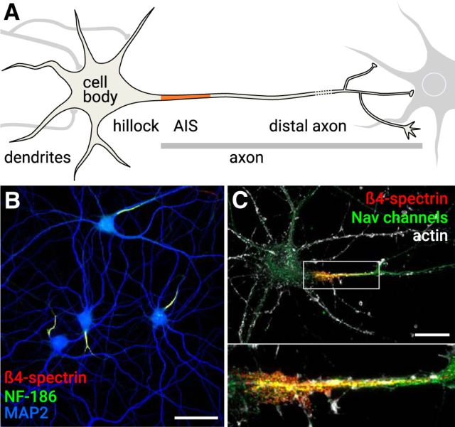
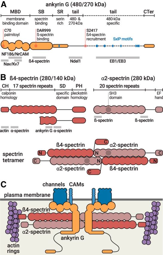
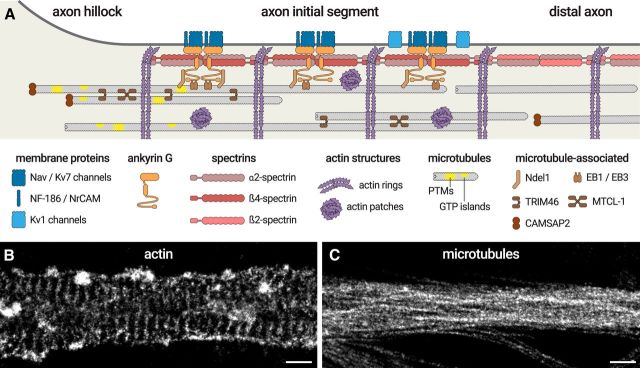
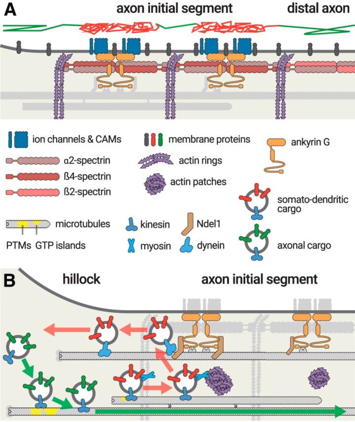
At the base of axons sits a unique compartment called the axon initial segment (AIS). The AIS generates and shapes the action potential before it is propagated along the axon. Neuronal excitability thus depends crucially on the AIS composition and position, and these adapt to developmental and physiological conditions. The AIS also demarcates the boundary between the somatodendritic and axonal compartments. Recent studies have brought insights into the molecular architecture of the AIS and how it regulates protein trafficking. This Viewpoints article summarizes current knowledge about the AIS and highlights future challenges in understanding this key actor of neuronal physiology.


Figures

Figure 1.
Figure 1.
The AIS. A, A typical neuron receives input on the cell body and dendrites (left). The hillock leads to the axon, which contains the AIS (orange). The distal axon contacts downstream neurons (right). B, Hippocampal neurons after 22 d in culture labeled for the AIS components NF-186 (green) and β4-spectrin (red). The somatodendritic compartment is labeled using an anti-MAP2 antibody (blue). Scale bar, 50 μm. C, Hippocampal neuron after 14 d in culture labeled for actin (gray), β4-spectrin (red), and Nav channels (green). Bottom, The zoomed image represents the AIS. Scale bar, 20 μm.
Figure 2.
Figure 2.
Molecular structure of ankyrin G and AIS spectrins. A, Domain organization of ankyrin G, which exists as isoforms of 480 and 270 kDa at the AIS (top, orange). Important residues (red) and EB-binding SxIP motifs (blue) are indicated. Binding sites of partners are indicated below the protein (gray bars). B, Domain organization of β4-spectrin, which exists as isoforms of 280 and 140 kDa (left), and domain organization of α2-spectrin (right). Binding sites of partners are indicated below the protein (gray bars). Bottom, Structure of the α2/β4 spectrin tetramer. N and C indicate the aminoterminus and carboxyterminus, respectively, of each subunit. C, The AIS submembrane complex. The α2/β4 spectrin tetramers (red) lie horizontally under the plasma membrane (dark gray), connecting actin rings (purple) with a distance of ∼190 nm. In the middle of the tetramer, ankyrin G (orange) is bound to β4-spectrin and anchors AIS membrane proteins (Nav/Kv7 channels, CAMs, blue).
Figure 3.
Figure 3.
The molecular organization of the AIS. A, Diagram depicting the organization of the AIS scaffold. Membrane proteins (Nav/Kv7 channels, NF-186, NrCAM, blue) are anchored by ankyrin G (orange), itself inserted into the α2/β4-spectrin complex (red) that spaces actin rings (purple). Kv1 channels (light blue) are present along the distal AIS. In the distal axon, the complex is made of α2- and β2-spectrin (pink). Ankyrin G binds to microtubules via EB1/EB3 proteins and Ndel1 (brown). AIS microtubules (gray) are capped by CAMSAP2 at their minus-end (brown) and grouped in fascicles crosslinked by TRIM46 and possibly MTCL-1 (brown). They are enriched in post-translational modifications (PTMs) and GTP islands (yellow). Intracellular patches of actin (purple) are also present inside the AIS. B, The <50 nm resolution of stochastic optical reconstruction microscopy (STORM) makes it possible to resolve the 190 nm spacing of actin rings and the presence of actin patches along the AIS. C, STORM can resolve microtubule bundles along the AIS, but the close apposition within bundles (∼30 nm spacing) makes it a challenge to distinguish individual microtubules. Scale bars: B, C, 0.5 μm.
Figure 4.
Figure 4.
Protein trafficking at the AIS. A, The AIS forms a diffusion barrier for membrane proteins and lipids (gray). A virtual trajectory (top) shows unimpaired diffusion at the cell body and distal axon (green segments), whereas diffusion at the AIS is limited by the AIS scaffold (red segments). B, Sorting of transport vesicles at the AIS entrance. Vesicles containing axonal cargoes (green) have kinesin motors (blue) that recognize cues on microtubules entering the axon, either post-translational modifications or GTP islands (yellow), before being transported into the axon (green arrow). Somatodendritic cargoes that start progressing into the AIS (red) are stopped by myosin-mediated immobilization at actin patches, before being brought back to the cell body by Ndel1-dependendant dynein transport (red arrows).

References

    1. Akhmanova A, Hoogenraad CC (2015) Microtubule minus-end-targeting proteins. Curr Biol 25:R162–R171. 10.1016/j.cub.2014.12.027 - DOI - PubMed
    1. Akin EJ, Solé L, Dib-Hajj SD, Waxman SG, Tamkun MM (2015) Preferential targeting of Nav1.6 voltage-gated Na+ channels to the axon initial segment during development. PLoS One 10:e0124397. 10.1371/journal.pone.0124397 - DOI - PMC - PubMed
    1. Al-Bassam S, Xu M, Wandless TJ, Arnold DB (2012) Differential trafficking of transport vesicles contributes to the localization of dendritic proteins. Cell Rep 2:89–100. 10.1016/j.celrep.2012.05.018 - DOI - PMC - PubMed
    1. Albrecht D, Winterflood CM, Sadeghi M, Tschager T, Noé F, Ewers H (2016) Nanoscopic compartmentalization of membrane protein motion at the axon initial segment. J Cell Biol 215:37–46. 10.1083/jcb.201603108 - DOI - PMC - PubMed
    1. Angelides KJ, Elmer LW, Loftus D, Elson E (1988) Distribution and lateral mobility of voltage-dependent sodium channels in neurons. J Cell Biol 106:1911–1925. 10.1083/jcb.106.6.1911 - DOI - PMC - PubMed